MR Renal Mass W/WO Protocol
Scan Notes
Last updated: 4/12/19
Charge as: Abdomen W/WO
Scanner preference: 1.5T
Coil: Torso Coil
Breath Holds:
- Scan on expiration.
- Monitor that patient is breath-holding. Breathe the patient slowly so they have time to follow instructions. Do not start scan until the patient has stopped breathing.
- Give 2L O2 if it will help with breath-holds UNLESS PATIENT HAS COPD OR ANOTHER REASON NOT TO GIVE O2.
- Send ADC maps and subtractions
| Plane | Weighting | Mode | Slice | Gap | FAT SAT | FOV | Notes |
|---|---|---|---|---|---|---|---|
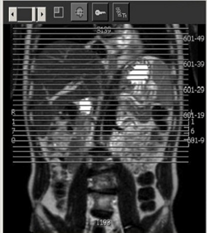
| Cor | T2 | SSFSE BH | 5mm | 0.5mm | No | Liver through bottom of kidneys | Ensure kidneys are well-centered in coil to ensure good signal at dome. |
| Axial | T1 | Dual Echo SPGR BH | 3mm | 0.5mm | No | Liver through bottom of kidneys | May be separated into overlapping stacks if patient cannot breath-hold. Do not interleave images. |
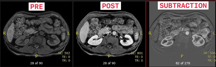
| Cor | T1 | 3D mDixon pre BH | 3mm | 0.5mm | No | Check before giving contrast. Minimize SENSE if there is mottling in the center of the image. Consider not using SENSE and allowing wrap into the peripheral image, but not into the kidneys | |
| Axial | T1 | 3D mDixon pre BH | - | - | Yes | Both kidneys | Check before giving contrast. Minimize SENSE if there is mottling in the center of the image. |
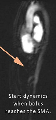
| Power Inject Contrast | Bolus Track | Trigger when bolus reaches the SMA | |||||
| Axial | T1 | 3D mDixon post BH x3 phases | - | - | Yes | Both kidneys | Check before giving contrast. Minimize SENSE if there is mottling in the center of the image. |
| COR | T1 | 3D mDixon post BH | - | - | Yes | Both kidneys | EXACT parameters as the COR mDixon precontrast. |
| Axial | T1 | 3D mDixon 4 minute post BH | - | - | Yes | Both kidneys | 4-minute post |
| Axial | T2 | TSE RT | 5mm | 1mm | SPAIR | Liver through bottom of kidneys | |
| Axial | T2 | DWI | 7mm | 1mm | SPIR | Liver through bottom of kidneys. | Trigger & track. Free-breathing sequence, so please position slices accordingly. |