MR Renal Arterial Sclerosis W/WO Protocol
Scan Notes
Last updated: 4/12/19
Charge as: Abdomen WWO
Scanner preference: 1.5T
Coil: Torso Coil
- See notes on positioning sat bands for Ax 3D BTFE
- Send ADC maps
- Send Subtractions
- FOV: do not include patient’s arms
Breath holds:
- Scan on expiration.
- Monitor that patient is breath-holding. Breathe the patient slowly so they have time to follow instructions. Do not start scan until the patient has stopped breathing.
- Give 2L O2 if it will help with breath-holds UNLESS PATIENT HAS COPD OR ANOTHER REASON NOT TO GIVE O2.
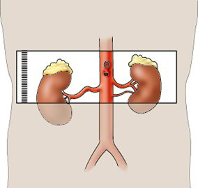
AX 3D BTFE SAT BAND NOTES
AX 3D BTFE: Total of 3 Sat bands
- 1 Oblique Sat band over each of the renal cortices, but NOT hila.
- Straight sagittal on the sagittal plane (aka straight coronal on the sagittal plane).
- Angled over renal cortices on transverse and coronal planes.
- 1 Oblique Sat band inferior to kidneys.
| Plane | Weighting | Mode | Slice | Gap | FAT SAT | FOV | Notes |
|---|---|---|---|---|---|---|---|
| Cor | T2 | SSFSE BH | 5mm | 0.5mm | No | Kidneys through pelvis | Ensure good signal over kidneys. |
| Axial | T1 | Dual Echo SPGR BH | 5mm | 0.5mm | No | Liver through kidneys | May be separated into overlapping stacks if patient cannot breath-hold. Do not interleave images. |
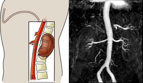
| Axial | T2 | 3D BTFE BH | - | - | Yes | Center over renal arteries | Three SAT bands. Please see NOTES and diagram below. |
| Cor | T1 | 3D mDixon pre BH | - | - | Yes | Kidneys through lower abdomen. Include the Aorta! | Ensure quality before contrast injection, ie wrap. Try not to use SENSE. Wrap into the edges of the image. Wrap cannot be in the image center (Thus, do not use SENSE).To decrease scan time, exclude anterior abdomen and paraspinal mucles. |
| Power Inject Contrast | Bolus Track | Trigger when bolus reaches the SMA | |||||
| Cor | T1 | 3D mDixon | - | - | Yes | Kidneys through lower abdomen. Include the Aorta! | Exact parameters as the pre-mDixon. Recon into thin axials & coronals. |
| Axial | T1 | 3D mDixon | - | - | Yes | Through renal arteries through bifurcation | May be done in 2 slabs. |
| Cor | T1 | 3D mDixon | - | - | Yes | Kidneys through lower abdomen | 5-minute post |
| Axial | T2 | TSE RT | 5mm | 1mm | SPAIR | Liver through kidneys | |
| Axial | T2 | SSFSE RT | 7mm | 1mm | Yes | Entire liver | Optional sequence if T2 SPAIR is poor quality. |