MR Liver Iron (Fe) Quantification WO BODY Protocol
Scan Notes
Last updated: 1/31/2024
Charge as: Abdomen WO
Scanner preference: MR2 or CHH2 only
Coil: Torso Coil
- No cardiac images to be acquired. ONLY LIVER images, please.
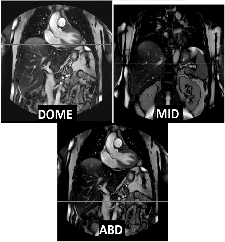
- Liver quantification consists of 3 sets of data, each sampling a different part of liver (dome, mid liver, inferior liver)
- The Fe sequences were created using a cardiac sequence, so you may have to put cardiac gating (either PPU or ECG) on the patient in order for proper gating.
- Send ADC maps
- Send Subtractions
- FOV: do not include patient’s arms
Breath Holds:
- Scan on expiration.
- Monitor that patient is breath-holding. Breathe the patient slowly so they have time to follow instructions. Do not start scan until the patient has stopped breathing.
- Give 2L O2 if it will help with breath-holds UNLESS PATIENT HAS COPD OR ANOTHER REASON NOT TO GIVE O2.
| Plane | Weighting | Mode | Slice | Gap | FAT SAT | FOV | Notes | |
|---|---|---|---|---|---|---|---|---|
| COR | T1 | 3D mDixon | - | - | Yes | Cover diaphragm to aortic bifurcation, abdominal wall to abdominal wall | ||
| Axial Liver Quantification(Dome) | T2* | FFE | 10mm | 10mm | None | Fit to Patient | One slice positioned at dome of liver. See images below | Breath Hold |
| Axial Liver Quantification(Mid-Liver) | T2* | FFE | 10mm | 10mm | None | Fit to Patient | One slice positioned at mid-liver. See images below. | Breath Hold |
| Axial Liver Quantification(Inferior liver/abd) | T2* | FFE | 10mm | 10mm | None | Fit to Patient | One slice positioned at lower portion of liver. See images below. | Breath Hold |