MR Adult Elbow WO Protocol
Scan notes:
- Add FABS sequences if clinical question is biceps pathology
- USE REST SLAB PROXIMAL TO ELBOW TO REDUCE FLOW AND PULSITILE MOTION
- If hardware is present:
- Do STIR instead of fat-sat mid-TE.
- If WITH CONTRAST, do non-fat-sat T1 post-contrast
Revised 11/21/2018
Charge as: Elbow WO
Scanner: 3T Only
Coil: Knee/Flex
| Plane | Weighting | Mode | Slice | Gap | FAT SAT | FOV | Scan Range |
|---|---|---|---|---|---|---|---|
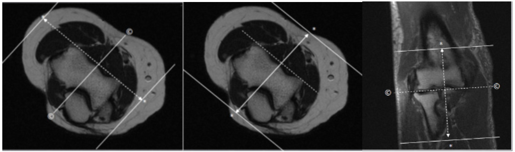
| AXIAL | PD | TSE | 3mm | 0.5mm | None | 14cm | 4cm Above/ Below Elbow Joint |
| AXIAL | Mid TE=40 T2 FAT SAT | TSE | 3mm | 0.5mm | SPAIR | 14cm | 4cm Above/ Below Elbow Joint |
| COR | T1 | TSE | 3mm | 0.5mm | None | 14cm | Angle to Distal Humeral Condyles |
| COR | T2 STIR (TE=40) | TSE | 3mm | 0.5mm | STIR | 14cm | Angle to Distal Humeral Condyles |
| SAG | Mid TE=40 T2 FAT SAT | TSE | 3mm | 0.5mm | SPAIR | 14cm | Angle to Distal Humeral Condyles |
| Optional FABS | Mid TE=40 T2 FAT SAT | TSE | 3mm | 0.5mm | SPAIR | 14cm | Perform if requested or if clinical question is biceps pathology.See info below for sequence set up. Call rad with questions. |
| Optional FABS | T1 | TSE | 3mm | 0.5mm | None | 14cm | Perform if requested or if clinical question is biceps pathology. See info below for sequence set up. Call rad with questions. |
Optional FABS elbow sequences
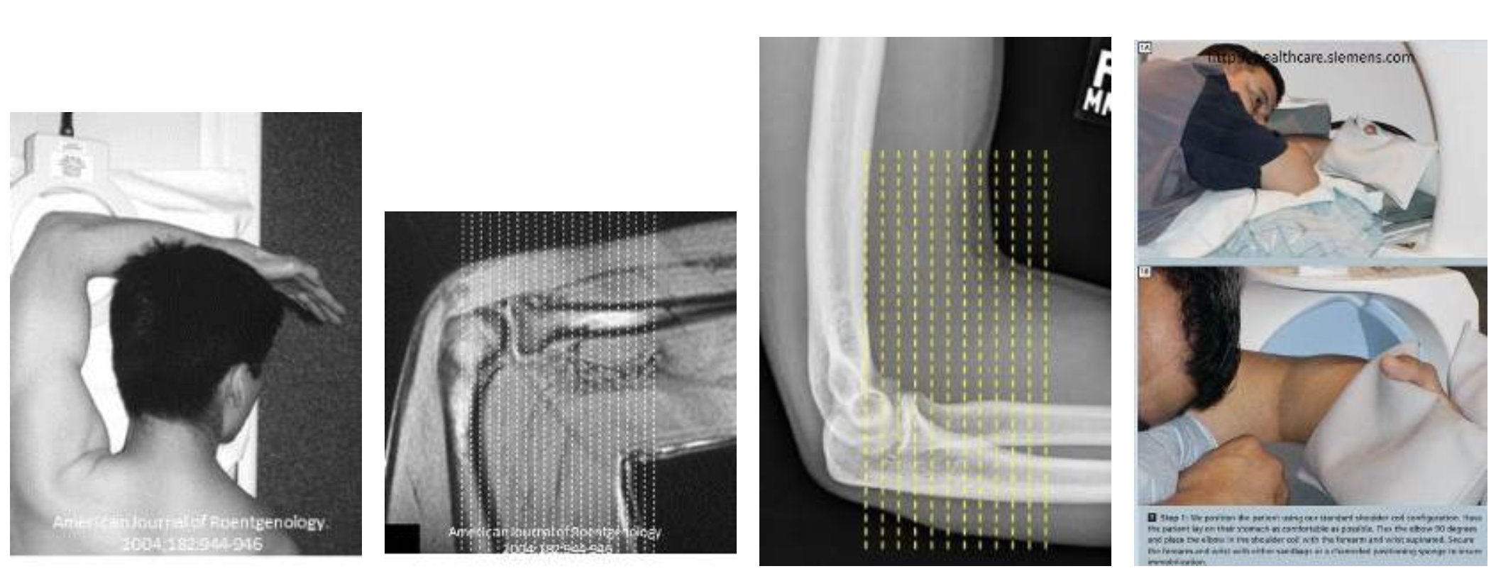
- Flexion, Abduction, Supination
- For better visualization of distal biceps tendon
- After usual sequences, place arm as shown (supination of wrist is important!)
- Images are oriented perpendicular to radius
- Images will be coronal to humerus but sagittal to rest of body