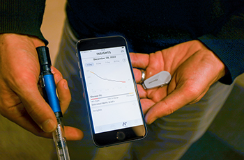
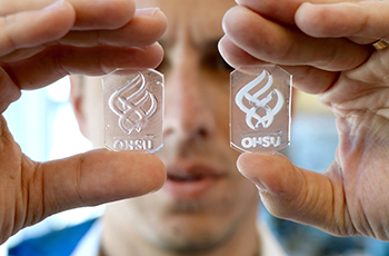
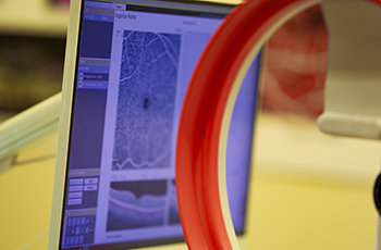
About the OCSSB

Joe Gray delivering a presentation on cancer biology

What is Systems Biology?

A global approach to biology

The study of medicine is changing from the old reductionist approach of looking at components of systems in isolation — individual cells or chemical pathways, for example — to a more global view of how the body works. The OHSU Center for Spatial Systems Biomedicine was established in 2011 as a joint project between the Knight Cancer Institute and the School of Medicine with this newer approach — called a constructionist, or systems biology approach — in mind. 

Multiscale means looking at all levels

We still want to measure and characterize DNA (genomics), RNA (transcriptomics) and protein profiles (proteomics)—to help us learn about differences between normal and diseased tissues, but we are gathering more information at both smaller and larger spatial scales — and watching this information as it changes over time. This multiscale systems biology approach, made possible through advances in measurement technologies, gives us much more context about what is happening in a healthy or unhealthy body. This, in turn, allows us to more fully understand how diseases develop, or, for example, how a normal, healthy person gets cancer. We believe that a spatial systems biomedicine approach will help us understand many areas of science and medicine. In particular, we are focused on solving how to prevent, treat, and cure cancers.

Leading technologies for multiscale research

At OCSSB, we have supported the development of many cutting-edge technologies to enable us to visualize cells directly, and obtain their omic profiles. Additionally, due to the extensive data collection that results from the imaging and omics analyses, the computer data server and storage requirements to support these are also necessary. Specific technological resources the OCSSB has contributed to include:

Learn more about these and other OHSU University Share Resources at the Research Cores web page, or read more about OCSSB Research projects and areas of interest. 

Videos

Our multiscale microscopy yields deeply detailed imaging data which can be composited into 3D or even four dimensions (3D images as they change over time). 

A single cancer "dragon" cell

This video is the product of many scanning electron micrographs composited to build the 3D image, subsequently manually edited to highlight the cancer cell (red). It demonstrates how a single cancer cell experiences diverse microenvironments because it physically spans such a large area, breaking out of normal cell confines.