People
Claudia López, Ph.D.
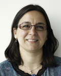
Claudia López Ph.D., director of the Multiscale Microscopy Core and research associate professor in the Biomedical Engineering Department, operationally oversees the Multiscale Microscopy Core. Dr. López has extensive experience with electron microscopy research including Transmission Electron Microscopy, Scanning Electron Microscopy, Dual-Beam applications and Serial Block Face Imaging. Her formal training is in biochemistry, biophysics and molecular biology and she has worked in the biochemistry-biophysics and microbiology fields for the past 22 years. Her expertise includes the analysis of both biological (cells, viruses, bacteria, particles and tissues) and non-biological materials (hard materials and fabrics) by TEM, SEM and FIB-SEM and she has routinely performed sample preparation (conventional and cryo-processing), electron microscopy imaging and image analysis. As part of her research interests Dr. López is developing correlative light and electron microscopy techniques for cultured cells and tissues, both for 2D and 3D electron microscopy. Dr. López is responsible for all day-to-day operations in the Multiscale Microscopy Core, including instrument maintenance and overseeing Multiscale Microscopy Core personnel; meeting with investigators to advise, plan and schedule new experiments; providing sample preparation and imaging services; training users; distributing data; explaining results, and billing for services.
Erin Stempinski, M.S.
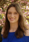
Erin Stempinski, M.S. is a research associate at the Multiscale Microscopy Core. She is experienced in conventional transmission and scanning electron microscopy, focused ion beam scanning electron microscopy, negative staining, gold immunolabeling of cryosections (Tokuyasu), and correlative light and electron microscopy methods. Erin has a background in botany and moved from working with plants to animals as an electron microscopy technician at the National Institutes of Health. She is responsible for sample preparation and imaging, training users, general lab maintenance and upkeep, and establishing new techniques. In addition to working in the Multiscale Microscopy Core, Erin supports research at the OHSU Center for Spatial Systems Biomedicine as part of the Gray lab.
Raakhee Shankar, Ph.D
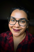
Raakhee Shankar, Ph.D. is an Associate Core Scientist at the Multiscale Microscopy Core. She is experienced in conventional transmission and scanning electron microscopy, electron tomography and segmentation, high pressure freezing and freeze substitution (rapid and AFS), negative staining, and gold immunolabeling on plastic sections. Raakhee’s training was mainly focused on cell and molecular biology, and she has worked with a variety of model organisms such as nematodes, mice, and cell culture. During her postdoctoral stint, she learned perfusion and sample preparation of rodent brain tissue at the National Center for Microscopy and Imaging Research. She is responsible for sample preparation and imaging, training users, general lab maintenance and upkeep.
Rodolfo (Rudy) Alvarado, M.S., C.E.M.T.
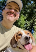
Rodolfo (Rudy) Alvarado, M.S., C.E.M.T. is an Associate Core Scientist in the Multiscale Microscopy Core at OHSU. He specializes in transmission electron microscopy (TEM), conventional room-temperature and cryo-scanning electron microscopy (SEM), serial block-face SEM (SBF-SEM), array tomography (AT), and cryo-electron microscopy workflows. His expertise also includes advanced biological sample preparation such as high pressure freezing/freeze substitution (HPF/FS), immunoEM, serial sectioning for TEM, single particle cryo-EM preparation and data acquisition, and 2D/3D image reconstruction and analysis. With a background in neuroscience and experience spanning from both biological and material sciences, Rudy brings over a decade of core facility experience to his role. Prior to joining OHSU, he served as a Core Research Facility Specialist at the University of Florida, where he supported diverse research projects and developed experience in confocal microscopy and correlative light and electron microscopy (CLEM). Across his career, he has worked with specimens ranging from nanoparticles to complex tissues and cellular systems across multiple model organisms. Within MMC, Rudy performs sample preparation and imaging, trains users on instrumentation, supports cryoEM data processing using platforms such as CryoSPARC Live on the Core’s Linux workstation (aka Hulk), and contributes to instrument troubleshooting and maintenance. In addition to his scientific work, Rudy is actively involved in OHSU’s Culturas Unidas Employee Resource Group (CUERG) as one of the leadership members. He also contributes to national microscopy standards through his service as a Certified Electron Microscopy Technologist (CEMT) and as a member of the Microscopy Society of America’s Certification Board, where he previously serviced as Chair.