DualBeam Scanning Electron Microscope
FEI Helios Nanolab™ G3 DualBeam™ Focused Ion Beam-Scanning Electron Microscope
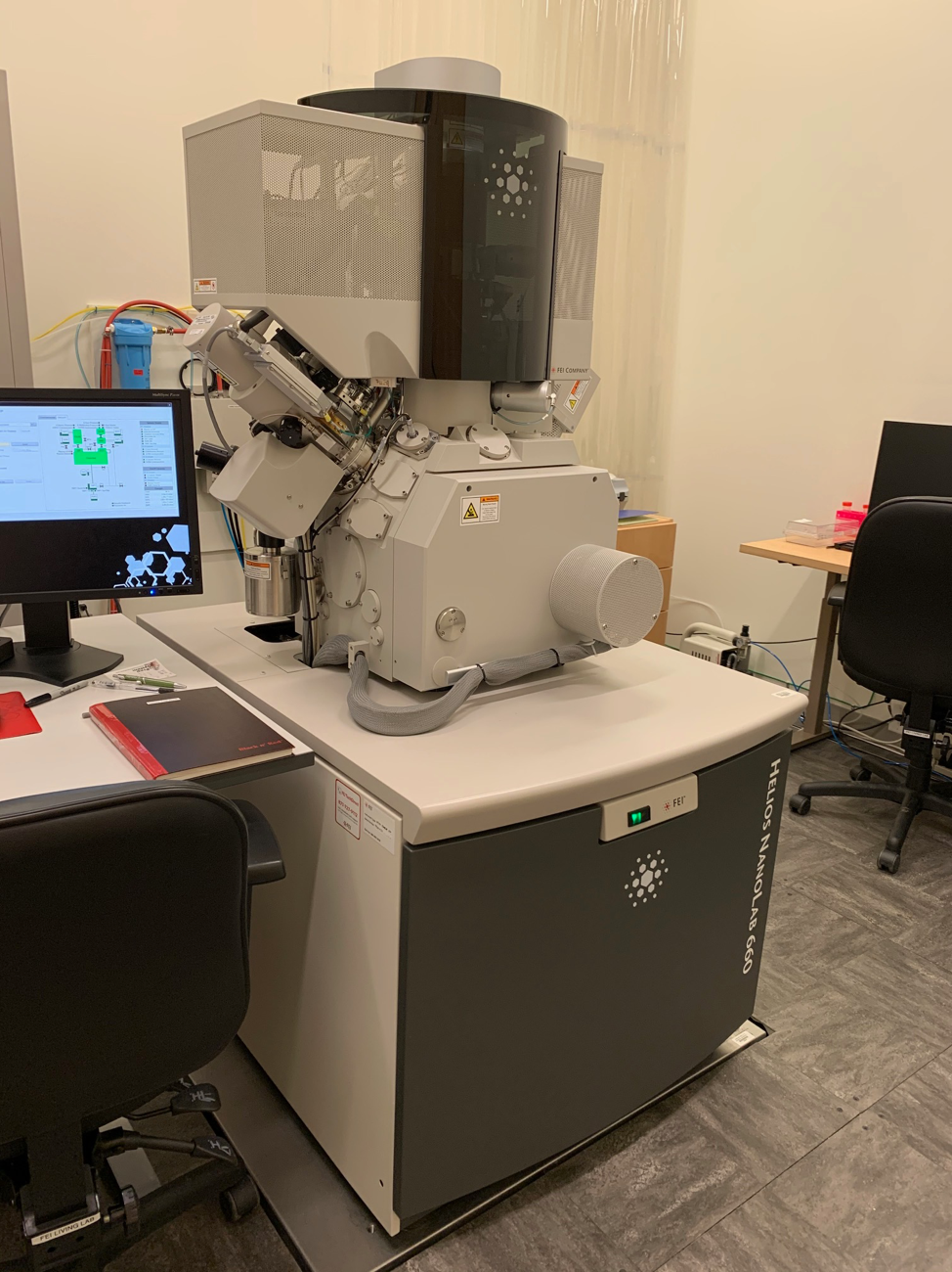
The FEI Helios Nanolab G3 DualBeam FIB-SEM platform is designed to access a new world of extreme high resolution 2D and 3D characterization. Precise FIB slicing, combined with a high precision piezo stage and superb SEM performance support automated software for high-resolution sample surface montaging, unattended sample preparation, or 3D characterization. Serial slicing and imaging using the AutoSlice and View™ software creates a 3D-image stack with 4-nm isotropic resolution that may be reconstructed in separate software for high resolution visualization. Traditional SEM imaging allows high-resolution detail of sample surfaces of all types and materials to be quickly and easily elucidated.
Features
- Ideal for the analysis of surface topographies or post-fixed resin-embedded samples by secondary or backscatter electron detection using the ETD, retractable CBS, and in-column TLD, MD, or ICD detectors.
- The focused gallium-ion beam ablates fine slices as thin as 4-nm to reveal structure under the resin block surface in order to provide a novel 3D picture of the sample volume.
- Maps™ software package allows large-format montaging over hundreds of micrometers, providing a “Google Street View”-like dataset.
- Imaging data from any outside source may be imported and correlated in the Maps software for Correlative Light and Electron Microscopy (CLEM)
- A retractable STEM detector allows low-voltage (10-30 keV) screening of TEM specimens.
Fast Facts
- Isotropic 3D data acquisition
- High Efficiency detectors for SE and BSE imaging
- Precision 5-axes motorized stage
- Better than 1.0 nm SEM resolution at all keVs
- Better than 4.0 nm FIB resolution at 30 keV
- Low kV FIB imaging down to 500 V
Location
- Robertson Life Science Building P2
Visit the ThermoFisher website for more information on the Helios
Publications featuring Helios images
Monique Y. Rennie, Curran G. Gahan, Claudia S. López, Kent L. Thornburg and Sandra Rugonyi. 3D Imaging of the Early Embryonic Chicken Heart with Focused Ion Beam Scanning Electron Microscopy. Microscopy and Microanalysis. 2014 Aug; 20(4):1111-9. doi:10.1017/S1431927614000828.