Helen Yang, Ph.D.
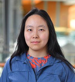
Assistant Scientist, Vollum Institute
Email: yanghe@ohsu.edu
Phone: 503-418-7552
Office: Vollum 4435A
Yang Lab (in progress)
Biography
Helen Yang earned her B.A. in Molecular and Cellular Biology from Harvard University in 2011. She then moved to Stanford University and earned her Ph.D. in Neurosciences in Tom Clandinin's lab, where she studied computations in the fruit fly early visual system using novel genetically encoded voltage indicators. She then did her postdoctoral training in Rachel Wilson's lab at Harvard Medical School, where she studied what features of walking are under the brain's control and where she was a key member of the team building the BANC connectome, a synapse-level wiring diagram of the adult female fruit fly central nervous system. Yang joined the Vollum Institute as an assistant scientist in 2026.
Summary of current research
The ability to actively move through the world is a defining characteristic of the animal kingdom. Locomotion underpins a wide range of essential behaviors, such as finding food, avoiding predators, and seeking out different environments. Critically, to support such a range of behaviors, locomotion is both reliable and flexible: animals can generate the appropriate locomotor movements when required and can produce a variety of locomotor outputs as conditions change. However, we do not understand the core principles that govern how nervous systems drive reliable, flexible locomotion. What circuit architectures support this? What information about the locomotor movement, the external world, or internal states is being processed in these circuits? The overall research goal of the Yang lab is to reveal how the nervous system generates reliable and flexible locomotion across timescales.
The Yang lab uses Drosophila walking as the model system for pursuing this goal, focusing on the neurons that carry signals between the brain and the nerve cord and that serve as a critical bottleneck in the neural control of walking. Like many vertebrates, fruit flies locomote through walking to accomplish various behavioral goals, and they face similar physical and neural challenges in the process. The Yang lab combines the rich genetic toolkit and the synapse-resolution wiring diagrams (connectomes) available in flies with detailed behavioral tracking, electrophysiology, two-photon imaging, and optogenetics to understand the neural circuit architecture, coding, and dynamics underpinning walking.
Selected preprints and publications
- Bates AS*, Phelps JS*, Kim M*, Yang HH*, ... Murthy M, Drugowitsch J, Wilson RI, Lee WA. (2026) Distributed control circuits across a brain-and-cord connectome. Nature, 656: 957-970.
- Pang MM, Chen F, Xie M, Druckmann S, Clandinin TR, and Yang HH. (2025) A recurrent neural circuit in Drosophila temporally sharpens visual inputs. Current Biology, 35(2): P333-346.E6.
- Yang HH, Brezovec BE, Serratosa Capdevila L, Vanderbeck QX, Adachi A, Mann RS, and Wilson RI. (2024) Fine-grained descending control of steering in walking Drosophila. Cell, 187(22): 6290-6308.e27.
- Isaacman-Beck J, Paik KC, Wienecke CFR, Yang HH, Fisher YE, Wang IE, Ishida IG, Maimon G, Wilson RI, and Clandinin TR. (2020) SPARC enables genetic manipulation of precise proportions of cells. Nat. Neurosci., 23: 1168-1175.
- Yang HH and Clandinin TR. (2018) Elementary Motion Detection in Drosophila: Algorithms and Mechanisms. Annu. Rev. Vis. Sci., 4:143-163.
- Chamberland S*, Yang HH*, Pan M, Evans SW, Chavarha M, Yang Y, Salesse C, Wu H, Wu JC, Clandinin TR, Toth K, Lin MZ, and St-Pierre F. (2017) Fast two-photon imaging of subcellular voltage dynamics in neuronal tissue with genetically encoded voltage indicators. eLife, 6:e25690, DOI: 10.7554/eLife.25690.
- Fisher YE*, Yang HH*, Issacman-Beck J, Xie M, Gohl DM, and Clandinin TR. (2017) FlpStop, a tool for conditional gene control in Drosophila. eLife, 6:e22279, DOI: 10.7554/eLife.22279.
- Yang HH and St-Pierre F. (2016) Genetically encoded voltage indicators: opportunities and challenges. J. Neurosci., 36(39):9977-9989.
- Yang HH*, St-Pierre F*, Sun X, Ding X, Lin MZ, and Clandinin TR. (2016) Subcellular imaging of voltage and calcium signals reveals neural processing in vivo. Cell, 166(1): 245-257.