People
Faculty
Andrew Adey, PhD, Molecular and Medical Genetics

Research: The Adey Lab is focused on the development of novel genomic and epigenomic technologies to produce a better understanding of the molecular regulatory networks that govern gene expression as it pertains to development and disease. These technologies are largely sequencing-based and performed at the single-cell level to enable the deconvolution of complex mixtures of cells of various cell types and states. We deploy these tools to probe the fundamentals of epigenetic control in the context of cancer, neurodevelopment, and neurodegeneration.
Techniques: We develop a number of technologies that produce genome-wide data that we use to uncover systems of gene regulatory control. This includes several single-cell technologies that profile chromatin accessibility (ATAC-seq), DNA methylation, chromatin folding, histone modifications, and other properties using a platform that can produce very large cell numbers and/or high coverage of single-cells; providing the requisite power to interrogate complex biological systems.
Teaching: Dr. Adey co-directs MGEN 625 Epigenetics & Reprogramming and is a lecturer in several courses where he focuses on genomic technologies and gene regulatory control (BME673, CANB616/CELL616, CANB622/MGEN622, BMSC662, BMSC664, CANB613/CELL613).
Peter Barr-Gillespie, PhD, Vollum Institute
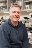
Research: Characterization of the development and function of the mechanotransduction mechanism of sensory hair cells.
Techniques: Transcriptomics (single cell and bulk), mass spectrometry (single cell and bulk), and quantitative immunocytochemistry.
Kimberly Beatty, PhD, Chemical Physiology and Biochemistry/Biomedical Engineering
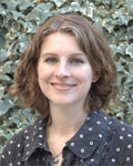
Research: The Beatty group at OHSU develops novel chemical tools and technologies for studying cellular proteins involved in human diseases.
Techniques: The Beatty group uses quantitative microscopy to understand how proteins organize and move in human cells
Teaching: Chemical Biology, BMSC 666
Steven Bedrick, PhD, Computer Science/Electrical Engineering
Research: My research interests are in applications of natural language processing to biomedical problems, in two main areas. First, in the analysis of biomedical text such as electronic medical notes, clinical trial descriptions, and scientific articles. The second area of interest is in the diagnosis and treatment of speech and language disorders, including post-stroke aphasia and Autism Spectrum Disorder.
Techniques: I use and develop a wide variety of machine learning models; of particular interest to me are novel language modeling architectures, embedding representations for heterogeneous (mixed structured, unstructured, and time-series) data, and domain adaptation methodologies.
Teaching: I teach courses in Natural Language Processing as well as Data Visualization.
Luiz Bertassoni, D.D.S, PhD, School of Dentistry
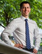
Research: Our research uses biomaterials engineering and biofabrication strategies (i.e. 3D bioprinting and organs on-a-chip) to address complex biological questions, mostly in calcified musculoskeletal and vascular tissues (i.e. craniofacial region, bone, bone marrow, and bone-affecting cancers). We also developed broad-range biofabrication tools, such as new bioprinting methods and organ on-a-chip platforms that address disease biology questions irrespective of the tissue in question. We have previously used these methods to address questions cancer metastasis, liver toxicity, brain development, and everything in between.
Techniques: Most of our work on quantitative and systems biology is focused on developing tools to mimic the complexity of native tissues. On that note, we develop and utilize methods that allow us to characterize and quantify the extend that our strategies allow us mimic these native systems, as well as other approaches that become necessary to understand cell behavior as a function of our ability to "re-create" them in-vitro. *Mostly experimental models. But we have used computation-assisted methods of quantification for the experimental models that we engineer, if that makes sense.
Teaching: Biocompatibility / BME 645
Young Hwan Chang, PhD, Biomedical Engineering
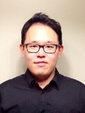
Research: My current research area aims to develop a general framework for quantifying multi-scaling imaging. Special emphasis will be on developing a novel image analysis tool to understand spatial distribution of cancer cells and their interactions. We use and adapt state-of-the-art machine learning/deep learning techniques for exploratory data analysis by rigorous data integration.
Techniques: We develop image analytics tools including segmentation, feature extraction and downstream analysis.
Teaching: I co-directed CONJ670 and BME674 (foundations/measurement science).
Siyu Chen, PhD, Ophthalmology & Biomedical Engineering
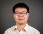
Research: My research aims to detect, differentiate and understand normal versus pathological features in the eye. My current projects focus on developing and applying new ophthalmic imaging technology to visualize the structure and function of the retina. We seek to investigate the connections between subcellular retinal morphology and visual function/impairment. Our hypothesis is that objective and quantifiable imaging biomarkers can facilitate both biomedical research and clinical management of eye diseases.
Techniques: We develop state-of-the-art ophthalmic imaging instruments and analytic algorithms to extract biomarkers for the diagnosis and monitoring of eye diseases. A specific interest is with optical coherence tomography (OCT), a depth resolved imaging modality that can generate three-dimensional structural and angiographic visualization of the eye.
Margaret Cheung, PhD, Pacific Northwest National Laboratory
Research: I am interested in the emergent properties of biomolecular interaction and organization using scalable computational tools and physical principles.
- Quantum mechanical calculations
- Atomistic and coarse-grained molecular dynamics simulations
- Mesoscopic simulations for active matter
Techniques: I develop analytical theories, computer algorithms, and mechanistic models that integrate the structural organization and collective dynamics of macromolecules across multiple time and length scales for the investigation of their emergent properties.
Teaching: Special Topics in Biological Physics (graduate Level), Statistical Mechanics (graduate level), Thermal Physics (upper undergraduate level)
Koei Chin, M.D., Ph.D., CEDAR/Knight Cancer Institute/BME

Research: The long-term goal of my research is to develop a strategy to improve therapeutic efficacy for cancer patients through: 1) understanding the mechanisms by which cancers are initiated and progressing, 2) early detection of high-risk cancer, and 3) understanding the mechanisms of therapeutic resistance at the level of organism, tissue and cell. To achieve this goal, we have focused on the technology development to quantify DNA abnormality, epigenetic state, molecular complexity, cell-to-cell interaction and tumor-microenvironment spatial interaction using fluorescently labeled probes. The developed cutting-edge technologies allow us to interrogate intracellular molecular architecture, cell state and spatial cell complexity in cancer tissues as systems biology approaches.
Techniques: We recently developed the Cyclic Immunofluorescence (CycIF) workflow for highly multiplexed immunostaining, in which a tissue is iteratively stained up to 60 proteins per assay for single cell phenotyping (PMID: 35243422). Also, we develop the multiplex image processing pipeline “mplexable” that is applicable to image data from a variety of imaging platforms such as CycIF, MICS, IMC, MIBI, etc. We use fluorescence in-situ hybridization (FISH) for DNA, RNA, and telomere quantification, which uncovered the highest genomic instability at the initiation of the malignant transformation (PMID: 15488760). Comparative genomic hybridization (CGH) has been used to interrogate gene copy number gain and loss in cancer genomes (PMID: 17157792 and 17157791). Our immunoFISH, the technology is superb to identify cell state with proteins and gene expression levels (PMID: 29184166).
Emek Demir, PhD, Molecular and Medical Genetics

Research: Our team specializes in integrating large scale -omic information and using them to answer cancer biology problems in conjunction with rich pathway information. Together with Chris Sander and Gary Bader, I led the development of BioPAX standard for pathway information and built an extensive software stack for aggregating and using all major public pathway data sources. Pathway Commons, our aggregation server, now has more than 2 million interactions and more than 400.000 highly detailed reactions of human pathways and it is the largest process-level pathway database. For the last 7 years, with the completion of key informatics components, I have shifted my focus to using this platform for answering cancer biology questions using statistics and machine learning.
Techniques: Our team developed multiple machine learning algorithms for detecting transcriptional impact of mutations, finding modulators of transcription factors, inferring differentially active networks from proteomic measurements and finding mutually exclusively altered pathway modules. As a part of two large DARPA projects - Big Mechanism and Communicating with Computers, we have also recently built tools for automating pathway curation and assembly through natural language processing and artificial intelligence. Our lab is increasingly focused on multi-omic modalities centered on proteomics and single cell sequencing.
David Dorr, M.D, M.S, Medical Informatics and Clinical Epidemiology, Chief Research Information Officer

Research: My research team, the Care Management Plus team studies the way data, information, and knowledge can help improve the health and well-being of vulnerable populations, including those with complex illnesses. We work on predictive algorithms, data integration, and pragmatic trials to understand the impact on outcomes. As Chief Research Information Officer, I am eager to support the infrastructure and capabilities needed to drive QSB efforts
Techniques: In my administrative role as Chief Research Information Officer, I work to support the tool ecosystem and am open to ideas and suggestions for how to expand this support and bring new capabilities into production.
Kyle Ellrott, PhD, Biomedical Engineering
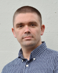
Research: Data science and data system engineering for cancer biology and genomics.
Techniques: Languages: Python, Go, R, Packages: Pandas, Matplotlib, ScikitLearn. Tools: Jupyter, docker, CWL, Galaxy Technologies developed: GRIP, a graph data integration and query system. BMEG, a large scale cancer genomics data integration project.
Teaching: BME 675
Andrew Emili, Ph.D.
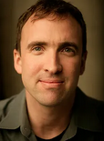
Research: My research program is centered on functional proteomics and systems biology-based investigations of biomolecular networks relevant to human health and development, and their disruption and rectification in disease states.
Techniques: Primarily precision mass spectrometry, but we favor integrative multidisciplinary team-based approaches.
Catherine G. Galbraith, PhD, Biomedical Engineering

Research: The two main project areas I am pursuing are: 1) Defining the cellular decision processes. — the decision modules and signaling pathways — that convert extracellular matrix probing into directed cell migration. 2) Capturing and correlating inhomegeneities in molecular behaviors with cellular functions.
Techniques: My work relies on cell biology, biophysics (mechanics), and microscopy. We concentrate on questions that have not been answered because they are technically challenging. We develop high risk/ high yield experiments paradigms and create custom analyses to quantify the data. Our approaches frequently rely on non-commercial experimental microscopy designs.
James K. Galbraith, PhD, Biomedical Engineering

Research: To visualize, quantify, and integrate the underlying molecular patterns responsible for cellular level functions such as migration, and synaptic plasticity
Techniques: Single molecule imaging, multimodal imaging, computer vision based image analysis and systems modeling.
Teaching: I am the lab director for the Review and Update of Neuroscience for Neurosurgeons (RUNN) course every year —I teach electrophysiology, biomechanics and single cell trauma models to neurosurgeons in a CME course.
Summer Gibbs, PhD, Biomedical Engineering
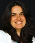
Research: The Gibbs laboratory is focused on novel fluorescence imaging technologies for microscopic and macroscopic cancer imaging. We have projects focused on fluorophore development to highlight tissues during fluorescence guided surgery and are developing fluorescence imaging technology to elucidate mechanism of cancer therapy response and resistance to targeted therapies.
Techniques: The Gibbs laboratory has expertise in small molecule fluorophore design and synthesis. We are also developing DNA barcoded antibodies for robust, nondestructive cyclic immunofluorescence imaging.
Teaching: Dr. Gibbs co-teaches BME608 – the qualifying exam preparation course twice annually. She also teaches BME669 – Physics of Medical Imaging.
Jeremy Goecks, PhD, Biomedical Engineering
Research: My laboratory’s current research program is focused on two broad areas: (i) computational infrastructure and machine learning approaches for biomedicine and (ii) precision oncology via integrative systems biology. These research areas are synergistic: the computational infrastructure and machine learning approaches that we develop is applied to our precision oncology efforts, and the analyses we want to do in precision oncology motivate the development of improved computational infrastructure and methods.
Techniques: The Galaxy computational workbench and Galaxy-ML toolkit, which we develop. Other tools we use include GSVA and VIPER for pathway/regulatory module activity levels, Scikit-learn and Keras for machine learning, and MCMicro for multiplex tissue image analysis.
Teaching: BME673, Cancer Systems Biology; BME605, Cancer Systems Biology Journal Club.
Visit the Goecks Lab's Website
Sara Gosline, PhD, Pacific Northwest National Laboratory
Research: Dr. Gosline is a computational biologist whose expertise focuses on data integration to employ a systems biology approach to rare disease and cancer. Specifically, she works to develop network algorithms in cancer and to translate these and other algorithms to more limited datasets that are available to study Neurofibromatosis Type 1, a rare disease that gives rise to numerous symptoms, most of which have no treatment.
Techniques: I primarily use statistical and basic machine learning models to integrate data and identify biomarkers of disease outcome. For analysis of sparse molecular datasets, I use graphical algorithms that collect data from published molecular interactions to build ‘networks’ of molecular interactions that can improve our understanding of these datasets.
Laura Heiser, PhD, Biomedical Engineering/Knight Cancer Institute
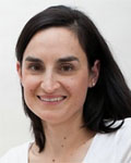
Research: My laboratory is focused on understanding the phenotypic and molecular responses of cancer and normal cells to diverse stimuli including small molecule inhibitors and growth factors. We use a variety of novel imaging-based and molecular techniques to assess dynamic changes in single cells, and have a particular interest in elucidating mechanisms of therapeutic response and resistance in cancer. In all of this work, we use well-integrated computational and experimental techniques with a key goal of closing the gap between these approaches.
Techniques: Experimental approaches include single-cell sequencing, immunofluorescence imaging, live-cell imaging, functional assays. Computational approaches include network analysis, clustering, and data integration.
Teaching: Introduction to Cancer Systems Biology (BME 673), Cancer Systems Biology Journal Club (BME 605)
David Huang, M.D., PhD, Ophthalmology/Biomedical Engineering
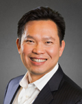
Research: My research interest is in optical imaging and laser therapeutics
Techniques: Our research group, the Center for Ophthalmic Optics & Lasers, develop optical coherence tomography (OCT) and OCT angiography for diagnostic imaging in the eye
Teaching: I teach in PHPH 621 Vision Science: Theoretical, Translational, and Clinical Aspects
Hiroshi Ishikawa, M.D, Ophthalmology
Research: My current research focus is AI in ophthalmology, which is heavily based on imaging data. I have been long working on medical image processing and clinical science related to glaucoma.
Techniques: I do not use any specific tool for my research. However, most of the data I process/handle are acquired using various medical imaging modalities (both commercial and prototype), such as optical coherence tomography (OCT), ultrasound biomicroscopy (UBM), fungus photography, etc.I use various deep learning models (2D/3D CNN, GAN, LSTM, etc.) for my clinical AI research.
Teaching: I give lectures based on my research a few times every year.
Peter G. Jacobs, PhD, Biomedical Engineering
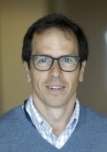
Research: My interests are in the area of medical device design, ubiquitous sensing and drug delivery technologies, machine learning, control systems, and signal processing as applied towards diabetes and other diseases. My lab is the artificial intelligence for medical systems (AIMS) lab where we develop new mathematical models of metabolism that are then integrated into control systems and decision support systems to help people with diabetes better manage their disease. One area of interest is how ordinary differential equation models of metabolism dynamics can be augmented by data driven models trained using machine learning.
Techniques: We propose different candidate model structures to describe metabolism dynamics, which requires use of a variety of tools in system identification including Bayesian inference methods such as Markov chain Monte Carlo and Hamiltonian Monte Carlo methods to solve for parameters in these candidate models. Part of our objective is to develop simulators of metabolism which mimic actual human metabolism. We have published details of these simulators and have made them open access for usage broadly around the world.
Teaching: I have taught BME 680 (Biomedical signal processing), BME 683 (Physiologic modeling and model predictive control), and BME 610NN (Bayesian data analysis). My lab also hosts a weekly machine learning and modeling reading group.
Yali Jia, PhD, Ophthalmology/Biomedical Engineering
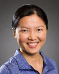
Research: My most active research areas are 1) technical development and clinical translation of OCT, OCT angiography, OCT oximetry; 2) functional OCT in animal models; 3) artificial intelligence to detect and classify retinal pathologies.
Techniques: Matlab (image processing), Python (AI), CUDA (GPU), and SPSS and MedCalc (statistics).
Teaching: I give one “optical coherence tomography” lecture in BME 674 Foundations of Measurement Science.
Yifan Jian, PhD, Ophthalmology
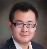
Research: My research interest is in optical imaging, high performance computing and artificial intelligence. My lab builds state of the art ocular imaging systems ranging from the cellular resolution adaptive optics system to widest possible field of view handheld imaging device.
Techniques: Lasers, Optical Engineering, High performance computing (parallel computing), AI, and imaging instrumentation.
Teaching: I have one “optical coherence tomography” lecture on BME 674 Foundations of Measurement Science.
James E. Korkola, PhD, Biomedical Engineering

Research: The Korkola lab studies how the tumor microenvironment impacts the behavior of cancer cells. We use innovative microarray technology to assess the impact of thousands of unique combinatorial microenvironment factors on cell phenotypes. We focus on how early tumor development is impacted by a pro-tumorigenic microenvironment (and in particular, the senescent microenvironment), as well as how therapeutic response is altered by microenvironmental factors.
Techniques: We use unique microenvironment microarray technology, which was developed in our lab, to study the tumor microenvironment in conjunction with advanced high-throughput imaging and image analysis approaches. We also make use of next generation sequencing (RNAseq, ssRNAseq, ATACseq) and protein analysis (Reverse Phase Protein Arrays) to understand transcriptional programs, expression signatures, and signaling pathway activation that are involved in response to microenvironmental perturbations and therapy.
Teaching: Dr. Korkola is the director of the Advanced Topics in Cancer course (CANB616; CELL616), and also lectures for the Cancer Systems Biology Course (BME673).
Ellen Langer, PhD, Molecular and Medical Genetics
Research: My primary research interest is to understand the molecular mechanisms underlying cellular plasticity during tumor initiation, response to therapy, and metastatic progression. Using 2D tissue culture, 3D bioprinted tissues, and mouse models, I am interrogating the cell-intrinsic and cell-extrinsic mechanisms by which tumor cells acquire or maintain the phenotypic heterogeneity that provides them with advantages in terms of growth, survival, drug resistance and metastasis.
Techniques: In order to assess the effects of intrinsic or extrinsic alterations to tumor phenotypes, I am developing manipulable, heterotypic 3D models of both breast and pancreatic cancer. To assess changes in cell state, I use many quantitative single cell approaches, including single cell RNA-sequencing, single cell ATAC-sequencing, and cyclic immunofluorescence image analysis.
Teaching: I teach a class on Metastasis for the CANB616 Advanced Topics in Cancer Biology course, and a class on Experimental Model Systems for the BME673 Cancer Systems Biology course
Bingbing Li, M.D., Ph.D., CPB
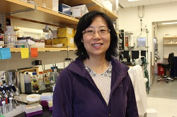
Research: The Li group at OHSU develops new tools for fundamental biological questions and new therapeutics for cancer patients. Her group is particularly interested in fusion-driven cancers and developing non-invasive imaging tools.
Techniques: The Li group implements quantitative pharmacology methods to develop small molecule tools and understand how small molecule drugs work in vitro and in vivo.
Teaching: PHPH 606-0
Sanjay Malhotra, PhD, Cell, Developmental and Cancer Biology
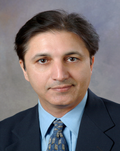
Research: My research interests focus on the design and discovery of synthetic and natural product inspired small molecules, which can provide insight into biological mechanisms and disease targets. The key aims of my research are (i) to design probe molecules that can be used to characterize the functions of particular proteins or drive specific cellular phenotypes, (ii) discover compounds that could be turned into drugs for human diseases and, (iii) educate the next generation of drug hunters.
Techniques: My laboratory employs the tools of synthetic medicinal chemistry, molecular modeling and chemical biology for translational research in drug discovery, development, imaging and radiation.
Clara Mosquera-Lopez, PhD, Biomedical Engineering
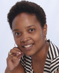
Research: My research interests lie at the intersection of biomedical signal processing and artificial intelligence for healthcare applications including digital health, data-driven modeling, development of technologies for diabetes management, and analysis of biosensors data. Currently, I work on the development of novel technologies for solving several glucose prediction problems including short-term hypoglycemia and hyperglycemia as well as nocturnal hypoglycemia in T1D, heart signals analysis, and fall detection and risk estimation using advanced machine learning techniques.
Techniques: I develop models and systems using Matlab/Python, particularly I used IA tools such as Tensorflow/Keras/Scikit-learn for creating machine learning pipelines. I use pandas/statmodels/pyMC for statistical analysis. At the AIMS Lab, we also use type 1 diabetes simulators developed in Matlab/Simulink for our in-silico testing.
Teaching: Currently, I do not teach any courses at OHSU. I lead a weekly machine learning reading group for AIMS Lab students, interns, scientists and faculty.
Learn more about the Artificial Intelligence for Medical Systems Lab
Xiaolin Nan, PhD, Biomedical Engineering/Knight Cancer Institute
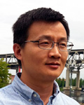
Research: Our current research focuses in three related areas: development of multiplexed fluorescence nanoscopy; multiscale spatial systems biology; and early cancer biology and detection.
Techniques: We study quantitative and systems biology using single-molecule and super-resolution microscopy in combination with chemical biology and computation.
Teaching: Our research topics are in part covered in CONJ 670: Foundations of Measurement Science, and in BME 645: Biocompatibility: Host-Implant Interactions.
Olga Nikolova, PhD, Division of Oncological Sciences
Research: My research focus is on graphical models for Big Data with applications to systems biology and personalized health, especially cancer. I am currently a Research Assistant Professor in the Division of Oncological Sciences, where I apply and develop new machine learning methods to help guide therapeutic strategies. I also lead the analysis of the first of its kind ex-vivo genome-wide CRISPR dataset, and its integration with the largest assembled dataset of matched multi-omic profiles, drug screens, and clinical information for primary leukemia samples from the Beat AML project. I also work in the space of single-cell data analysis and integration.
Techniques: I mostly use Bayesian predictive models to model drug response in patient samples. I have developed most of the models and workflows I use, e.g. the Gene-wise prior Bayesian Group Factor Analysis Model, various parallel algorithms for Bayesian network structure learning, siRNA and CRISPR screen data analysis. (Please let me know if you would like publication references.)
Vladislav Peytuk, PhD, Pacific Northwest National Laboratory
Research: Dr. Petyuk’s research interests are applying quantitative LC-MS proteomics and development of data analysis tools. The primary applications are focused on neurodegenerative disorders and aging. He is particularly interested in the role of insulin and insulin-like signaling in Alzheimer’s disease and cognitive decline.
Techniques:
- Quantitative LC-MS proteomics
- R/Bioconductor packages for processing proteomics data
- CRNT4SBML, a tool for assessing bistability within signaling pathways
Carmem Pfeifer DDS, PhD, School of Dentistry
Research: biomaterials, polymer materials, organic synthesis, antimicrobial materials, high strength materials, degradation-resistant materials.
Techniques: IR spectroscopy, NMR spectroscopy, universal mechanical testing, dynamic mechanical analysis, rheology (including biofilms and tissues), GC-MS, HPLC, microbiology assays (crystal violet, luciferase assay, EPS production assays), enzyme activity assays (zymography - gel and in situ, dynamic light scattering), SEM and confocal imaging.
Teaching: Dental Materials (at SOD) for first- and third-year dental students; introduction to polymer science (graduate students in Dental Materials). DM 711: Introduction to Dental Materials (Direct restorative materials) - DS1
DM 712: Introduction to Dental Materials (Indirect restorative materials) - DS1
DM 731: Clinical applications of dental materials - DS3
FOP - 511: Introduction to polymers in dental materials science (graduate students at Piracicaba School of Dentistry, Brazil)
Steve L. Reichow, PhD, Portland State University
Research: We are experts in a revolutionizing imaging technology known as cryo-electron microscopy (Cryo-EM), which we use to illuminate the 3D-structures of biological macromolecules and characterize their functional and disease-state mechanisms with atomic-level detail.
Technique: We apply cutting-edge methods in the high-resolution imaging technology of cryo-electron microscopy (Cryo-EM), together with biochemical, biophysical and computational tools to unveil the inner-workings of individual protein molecules, nature’s nano-machines. It is our hope that the atomic-level models and mechanistic insights developed by our research will help to elucidate the biomolecular basis of human health and disease.
Teaching: Biochemistry (CH591); Protein Structure and Dynamics (CH598); Biophysical Methods in Structural Biology (CH546) (taught at PSU)
Visit the Reichow Laboratory of Biomolecular Cryo-EM Website
Sandra Rugonyi, PhD, Biomedical Engineering
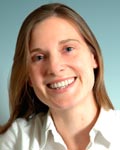
Research: Dr. Rugonyi’s research is on cardiovascular mechanics, with a focus on quantifying cardiovascular blood flow dynamics (hemodynamics) and understanding how mechanical stimuli from blood flow affects cardiovascular tissues, from embryonic development to adulthood. When blood flow is perturbed during embryonic development, congenital cardiac defects and vascular anomalies ensue; and anomalous blood flow conditions in adulthood lead to cardiovascular disease progression and pathophysiology, including aneurysms, and blood clotting/thrombosis. The details of how mechanical feedback from blood flow occurs, however, is largely unknown, and Dr. Rugonyi’s research aims at unravelling mechanisms and functional relationships that describe hemodynamic behaviour and how blood flow affects cardiovascular tissues, to ultimately predict risks and outcomes of cardiovascular interventions.
Techniques: Dr. Rugonyi’s research lies at the interface of Mechanics, Biology and Medicine. Projects include a combination of data acquisition (e.g. live imaging, digital data, 'omics' data), data analysis and quantification (e.g. image analysis, bio-statistics, trend quantification, artificial intelligence), software and algorithm development (e.g. computer vision, predictive modeling, uncertainty quantification). Importantly, Dr. Rugonyi’s group has developed computational fluid dynamics (CFD) models of blood flow under diverse conditions, such as during heart development, aneurysm formation, and cardiovascular interventions.
Teaching: BME 640: “Fluid Mechanics and Biotransport”. The course covers basic physical principles that determine the behavior of fluids, and the equations that describe such behavior.
Arpiar Saunders, PhD, Vollum Institute
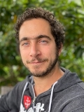
Research: Developmental and degenerative disorders alter the brain in largely unknown ways.
Our aims in research are focused on systematically mapping the complex cellular organization of the brain in high-throughput by developing and applying apply single-cell, single-virion genomic technologies. We are also interested in understanding the mechanisms through which viruses interact with diverse brain cell types.
Techniques: Our lab combines molecular engineering and computational approaches to collect and analyze genome-wide molecular measurements from hundreds of thousands of individual cells. We engineer “molecular barcodes” into viral genomes to track molecules derived from individual virion. We develop novel computational tools to analyze our datasets.
Teaching: I participate in the Neuroscience Graduate Program Bootcamp.
Carsten Schultz, PhD, Chemical Physiology and Biochemistry
Research: The Schultz group is interested in monitoring and manipulating intracellular signaling networks with a focus on alpha and beta cell biology, growth factor signaling and lung inflammation. We develop new genetically encoded and small-molecule-based fluorescent reporters and sensors to follow events in real time. We employ quantitative imaging of signaling events such as calcium and cAMP transients and oscillations and frequently monitor translocation between intracellular membrane systems. For image analysis, we employ ImageJ and FluoQ, a FIJI-based set of macros designed to rapidly provide presentable data, developed in our lab.
Teaching: I teach in the PBMS course “Chemical Biology Innovators” (BMSC 669).
Ujwal Shinde, PhD, Chemical Physiology and Biochemistry/ Knight Cancer Institute
Research: In the Shinde Laboratory research focuses on understanding the structure, folding, and evolution of proteins including proteases, kinases, and GPCRs and in particular, how the interactions and dynamics of these proteins can regulate their biological functions. The lab employs biophysical, biochemical and computational approaches to address these questions.
Techniques: We are currently not developing any tools used in quantitative and systems biology
Teaching: Courses taught are:
- BMSC 661 Structure and Function of Biomolecules
- BMSC 668 Molecular Biophysics and Experimental Bioinformatics
- BMB Journal Club: Protein Structure and Function
Charles Springer, PhD, Advanced Imaging Research Center

Research: We develop new forms of in vivo MRI. Most recently, we have introduced Activity MRI [aMRI]. With it, we have found non-invasive diffusion-weighted MRI can do the job of ubiquitous 18FDG-PET but with: (a) significantly higher spatial resolution, (b) considerably less cost, and (c) no pharmacokinetic requirement – it can be repeated continually.
Techniques: In AIRC, we have a wide range of in vivo MR instruments – ranging from ultra-high field human and small animal systems to instruments for biological tissue samples. Typically, we innovate new ways of acquiring data from these.
Teaching: In 18 years at OHSU, I have taught in the CONJ 670 [Foundations of Measurement Science], CONJ 671 [Analysis in Quantitative Bioscience], CELL/CANB 616 [Advanced Topics in Cancer Biology], and CH410/510 [NMR Spectroscopy] courses. In 35 years on the Stony Brook and Brookhaven National Laboratory Chemistry faculties, I taught in 16 different courses – ranging from first-year General Chemistry to Graduate level courses in magnetic resonance and inorganic chemistry. I also taught in Biochemistry and Physiology Department courses. In 2017, I received the Outstanding Teacher Award from the International Society for Magnetic Resonance.
Xubo Song, PhD, Medical Informatics and Clinical Epidemiology/Computer Science and Engineering Graduate Program
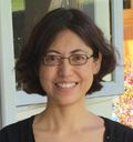
Research: My research focuses on two goals: (1) to develop novel computational methodologies to advance the fields of machine (deep) learning and computer vision, and (2) to apply these methodologies to the biomedical domain for computer-aided diagnosis, therapy, and decision support, through a multidisciplinary team-based approach.
Techniques: We develop quantitative and system biology tools to help researchers addressing basic science and clinical questions. These tools include image processing and analysis, machine learning/deep learning, and time series analysis.
Teaching: I teach three courses: (1) Machine Learning, (2) Advanced Topics in Machine Learning, and (3) Introduction to Image Processing. BMI543/643
Ou Tan, PhD, Casey Eye Institute/Biomedical Engineering
Research: Diagnosis and prediction of glaucoma and dementia using machine learning and ophthalmic imaging, especially optical coherence tomography (OCT)
Techniques: Matlab; based on this platform, I developed software for OCT image analysis and retinal blood flow measurement
Guillaume Thibault, PhD, Biomedical Engineering
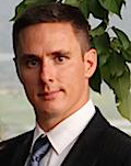
Research: My work interest focuses on developing tools and pipelines to help biologists and doctors to have a better understanding of their data. These tools are applied to a large variety of imaging modalities like H&E, cyclic immunofluorescence, MRI, and electron microscopy, and include all the crucial steps of analysis starting with registration, segmentation, features extraction until data analysis.
Reid F. Thompson, M.D, PhD, Radiation Medicine/Biomedical Engineering
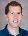
Research: My research interests include immunotherapy, genomics, clinical data science, precision medicine, and artificial intelligence in medical imaging.
Techniques: We develop novel tools predicting antigen processing, presentation, and response.
Zheng Xia, PhD, Biomedical Engineering
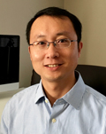
Research: We are a data-driven computational biology laboratory. We develop bioinformatics tools for next-generation sequencing data analysis and Machine Learning algorithms for large-scale biomedical data interpretation. Our current research interests include single-cell data analysis, machine learning prediction models for big data analysis, and omics data mining in cancer immunotherapy.
Techniques: Regulon analysis, Pathway enrichment tools, Cell type identification
Teaching: BME675
Craig K. Yoshioka, PhD, Biomedical Engineering/PNCC
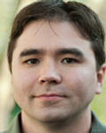
Research: Most interested in R&D for cryo-EM tools, traditionally software tools, but also in designing operational and organizational infrastructures/facilities, like the national cryo-EM center. Collaborating with other PIs to help interpret protein structure dynamics from cryo-EM experiments. Fairly comfortable with a wide-swath of biological systems; past collaborations include cryo-EM of viruses, ion channels, transporters, nucleosomes, ribosomes, etc.
Techniques: Pretty much everything in the single-particle cryo-EM workflow, but also somewhat familiar with cryo-electron tomography. I do not (currently) develop any of the components used in this work, but am always at the cutting-edge of pushing the latest tools and advancements to, and past, their limits.
Teaching: As part of the national cryo-EM center, I help organize and teach several workshops on cryo-EM (for conferences and independently), including being the founding member of an NIH-sponsored initiative to create a “merit badge” (certification) program for practical skills in cryo-EM workflows.
Daniel Zuckerman, PhD, Biomedical Engineering
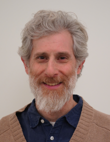
Research: The Zuckerman group uses physics-based computational methods to explore a range of problems from molecular to cellular scales, working extensively with experimental and theory collaborators. We are particularly interested in non-equilibrium processes such as occur in cell-surface transporters and other molecular machines, as well as in whole-cell morphodynamics. We also study flexible, dynamic systems such as multi-protein complexes using simulation and microscopy analysis tools. Because the laws of statistical physics govern molecular and celullar behavior, it is essential to apply physical tools to understand biology at these scales.
Techniques: The group uses and develops a wide range of methods. We develop specialized molecular dynamics (MD) algorithms to enable sampling of challenging processes such as protein-ligand (un)binding, protein folding and conformational changes. We also develop physically based analyses of the large data sets resulting from biomolecular MD. We employ kinetic (differential equation) models of molecular and cellular processes, in conjunction with Bayesian inference of parameters from experimental data. We analyze microscopy data, ranging from live-cell to electron microscopy, developing novel tools as needed.
Teaching: BME 675 (Analysis of Quantitative Bioscience) and BME 673 (Cancer Systems Biology)
Executive Committee - Quantitative and Systems Biology Program
Kimberly Beatty, Ph.D.
Summer Gibbs, Ph.D.
Laura Heiser, Ph.D.
Peter Jacobs, Ph.D.
Planning Committee - Quantitative and Systems Biology Program
Steven Bedrick, Ph.D. - Department of Medical Informatics and Clinical Epidemiology
Yifan Jian, Ph.D. - Department of Ophthalmology, COOL Laboratory
Bingbing Li, M.D., Ph.D. - Department of Chemical Physiology and Biochemistry
Clara Mosquera-Lopez, Ph.D. - Department of Biomedical Engineering
Olga Nikolova, Ph.D. - Division of Oncological Sciences
Arpiar Saunders, Ph.D. - Vollum Institute
Contacts - Quantitative and Systems Biology Program
Daniel M. Zuckerman, Ph.D., Director zuckermd@ohsu.edu
Caitlin Currie, Administrator qsb@ohsu.edu