Transmission Electron Microscopes
FEI Tecnai™ with iCorr™ (Integrated Light and Transmission Electron Microscope)
Description of the FEI Tecnai™ with iCORR™
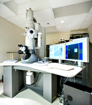
The most critical "tie point" between imaging modalities is between fluorescence and electron microscopy, with electron microscopy providing Angstrom to nanometer resolution information about molecular ultrastructure and FM providing nanometer to micrometer resolution information about functional molecular complexes. Scientists from FEI and the Biomolecular Imaging program at Utrecht University have developed novel sample fixation and staining procedures for the Tecnai™ with iCorr™ that preserve both ultrastructural and molecular features for fluorescence microscopy and transmission electron microscopy and reduce sample preparation times.
Location
Robertson Life Sciences Building
Online
November 2012
Fast facts
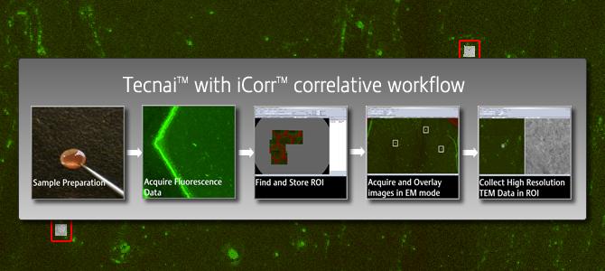
- Correlates light microscopy and electron microscopy. Uses light microscopy to find a rare cell or event and uses electron microscopy to study that event.
- Ideal for identifying rare cells, events in a large sample, such as 1 in 10,000 events or cells.
Features
- Locate precise regions of interest quickly
- Wide field excitation (470-490 nm)
- Optimized for green fluorescence
- Objective lens (NA 0.5, 0.65 um res)
- 5 Mpixel CCD
- Fluorescence and reflection mode
- Software controlled
More information
Glacios Cryo-Electron Microscope with Gatan K3 Camera
About the Glacios
The Glacios Cryo Transmission Electron Microscope (Cryo-TEM) from Thermofischer delivers a complete and affordable Cryo-EM solution to a broad range of scientists. It features 200 kV XFEG optics, the industry-leading Autoloader (cryogenic sample manipulation robot), and the same innovative automation for ease of use as on the Krios G4 Cryo-TEM. The Glacios Cryo-TEM bundles all this into a small footprint that simplifies installation.
The Glacios microscope will be equipped with the Gatan K3 camera, the new imaging performance benchmark for direct detection cameras. This next generation camera is optimized for the most demanding low-dose applications in both life science and materials science research. The K3 camera is the complete and latest expression of Gatan's deep experience in the delivery of real-time, single electron counting direct detection cameras.
Gatan K3 camera Features:
- Powerful inline signal processing will raise the DQE beyond that of the K2 camera
- Optional inline, GPU-based motion correction avoids the need to save terabytes of raw frames
- 24 megapixels (5,760 x 4,092) field of view – 1.6 times the size of the K2 camera
- 1,500 full frames per second – 3.75 times the speed of the K2 camera
Visit the ThermoFischer webpage describing the Glacios system
Visit the Gatan webpage describing the K3 camera system
Download the mag/pixel size table for the K3
Location:
Robertson Life Science Building P2