Our research projects
We are currently working on numerous collaborative studies involving different host pathogen interactions, models of inflammation, immunity, etc. in rhesus, cynomolgus and Japanese macaques. Here at OHSU we are collaborating with the Picker, Estes, Hansen, Okoye, Wilder, Axthelm, Wong, Streblow, Hirsch, Defilippis, Skalsky, Burwitz, Kievit, Jensen, Hessell, and Sacha labs and more details on our collaborative work can be found on their lab pages. We have numerous offsite collaborators as well with work in contraceptives, cardiac imaging, as well as infectious diseases and inflammation.
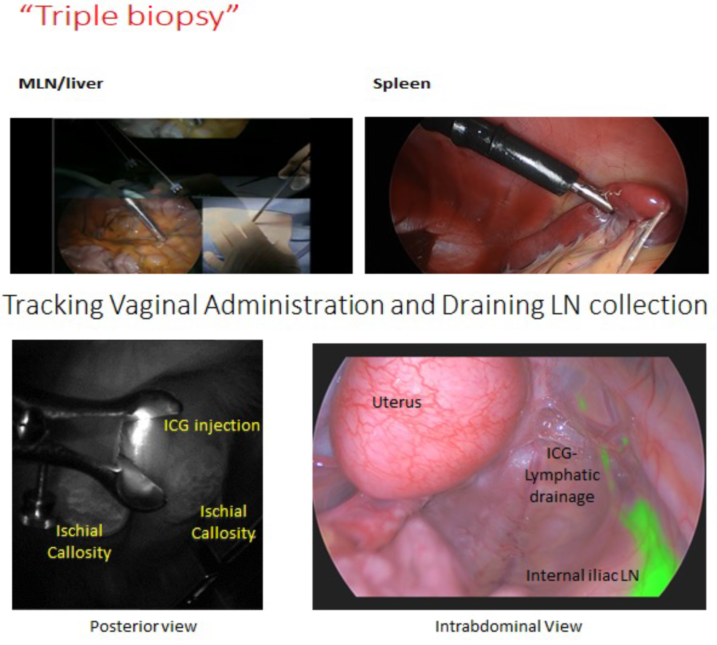
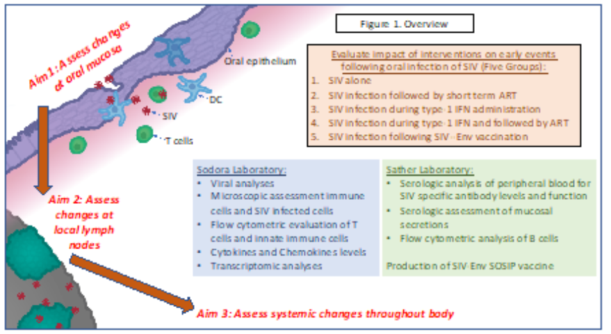
Our current research focuses on early events associated with viral dissemination from the initial site of infection and reservoir establishment in SIV macaque models. We have developed methods for establishing a localized infection and tracking that infection to the local draining lymph nodes using fluorescence imaging and systemically using our minimally invasive biopsy techniques for spleen and mesenteric lymph nodes.
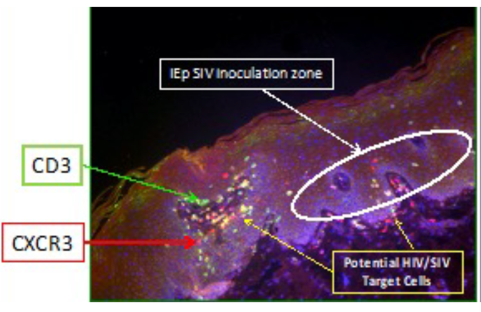
To accomplish this we will use an intraepithelial infection route in the oral mucosa (intraepethial or IEp injection).
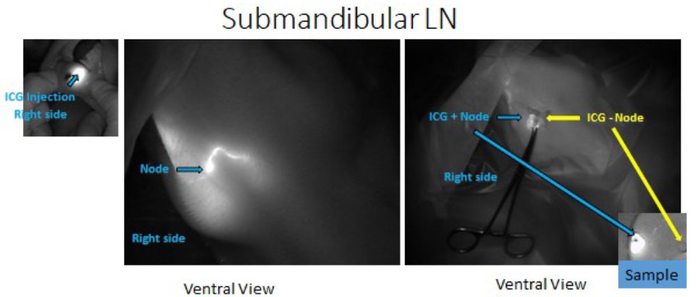
We will then follow the virus to the local draining submandibular nodes using NIR imaging.
We can evaluate early interventions such at antiretroviral therapy, vaccines, etc on viral dissemination kinetics to the local draining nodes, spleen, MLN, as well as how these impact the early viral host interactions that eventually determine the viral setpoints and reservoir sizes.
We are also continuing to improve our comprehensive sampling techniques optimizing sample quality, animal welfare, and frequency/number of timepoints to best answer experimental questions in macaque models. Current work involves further laparoscopic techniques for guided sampling of lymph nodes, liver partial lobectomies, and sampling of additional abdominal nodes.