Projects
How do you make an astrocyte, and what does it do?
Astrocytes are the most abundant cell type in the mammalian brain. We made the recent exciting discovery that astrocytes are also present in the Drosophila brain and are now using the fly to explore how these cells develop, the roles they play in neural circuit formation, and how astrocytes modulate brain function and behavior.
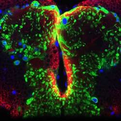
Image caption: A single astrocyte (green) in the larval central nervous system.
How do glial cells recognize and dispose of neuronal debris?
During normal development or after nervous system injury or disease, neuronal processes (axons, dendrites, and synapses) can degenerate and neuronal cell bodies often undergo apoptotic cell death. Glial cells are the primary cell type responsible for recognizing and clearing this neuronal debris. We are interested in understanding how neurons signal to glia to indicate debris is present and needs clearance, and the molecular basis of glial recognition and phagocytosis of neuronal debris.
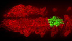
Image caption: Astrocytes (green) at metamorphosis transform into phagocytes to prune neurons.
How are long axons wrapped and supported by glia?
Long axons in mammals and flies are surrounded, and often individually ensheathed, by glial processes. Such insulation is thought to be critical for enhanced nerve conduction velocity and trophic support of long axons that are some distance from the cell body. We are exploring the molecular basis of axonal ensheathment in Drosophila and the mechanisms by which surrounding glial cells promote the survival and function of the axons they ensheath.
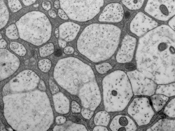
Image caption: EM cross-section of the larval nerve showing glial membranes (dark gray) surrounding axons.
How do axons undergo auto-destruction?
Severed axons (and dendrites) degenerate after axotomy, but is this a passive wasting away or an active death process? We recently discovered that deletion of the dSarm/Sarm1 gene resulted in the long-term survival of the distal portions of severed axon in both flies and mice. This work provided direct evidence for the existence of a dSarm/Sarm1-dependent axon death signaling pathway. We are now using powerful molecular approaches in Drosophila to identify additional axon death genes, and exploring the role of axon death signaling in neuron loss during disease using both fly and mouse models of neurological disorders.
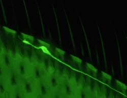
Image caption: A single sensory neuron in the adult Drosophila wing.