Neuroimaging Laboratory
The Neuroimaging Laboratory at the Layton Oregon Aging and Alzheimer's Disease Research Center (OADC) was established in 1989. Since then it has maintained a research focus centered on detecting early brain changes associated with cognitive decline and dementia. The lab manages the neuroimaging component of all studies at the Center.
In addition to acquisition and archival services, the lab serves as an imaging analysis resource specializing in volumetric analysis, white matter integrity and blood flow quantification. Additional assistance including statistical analysis and preparation of materials for presentation and publication is available.
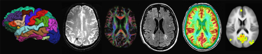
The Neuroimaging Lab conducts MRI studies on both 3 and 7T MRI systems using advanced sequences, employing a multimodal approach to brain imaging research. The Layton Center also manages a library of thousands of digitized MRI scans, including what is believed to be the largest collection of longitudinal MRI scans of cognitively intact elderly subjects.
For more information please contact:
David Lahna
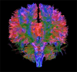
human diffusion imaging
Research projects
-
The OADC Neuroimaging lab collects and analyzes longitudinal MRI of healthy aging people with the goal of detecting brain changes that may predict cognitive decline. We measure cerebral blood flow, white matter microstructural integrity and structural volumetric changes. A major emphasis of the lab is white matter hyperintensities, which are a marker of vascular brain health in older individuals.
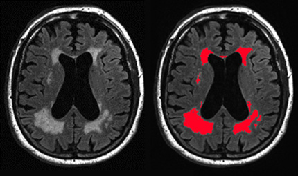
Automated white matter hyperintensity segmentation
We use a multimodal imaging approach to examine changes in perfusion and white matter integrity surrounding white matter hyperintensities. Changes in these measures in the cerebral blood flow penumbra predict expansion of hyperintensities and help us to understand the mechanisms for their growth.
Layers surrounding white matter hyperintensities where decreased blood flow predicts future expansion over time
-
We have developed a protocol for imaging postmortem brain tissue hemispheres and slices at 7 tesla strength MRI. The high-resolution images we collect are coregistered to in vivo images and used for MRI guided histopathological sampling. This allows us to examine MRI observed pathology at the microscopic level to better understand disease processes related to aging, vascular disease and dementia.
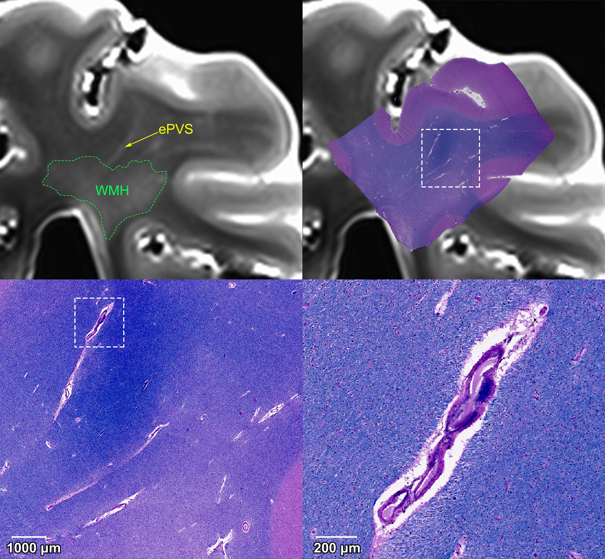
White matter hyperintensities and enlarged perivascular spaces identified in postmortem MRI are sampled histologically. 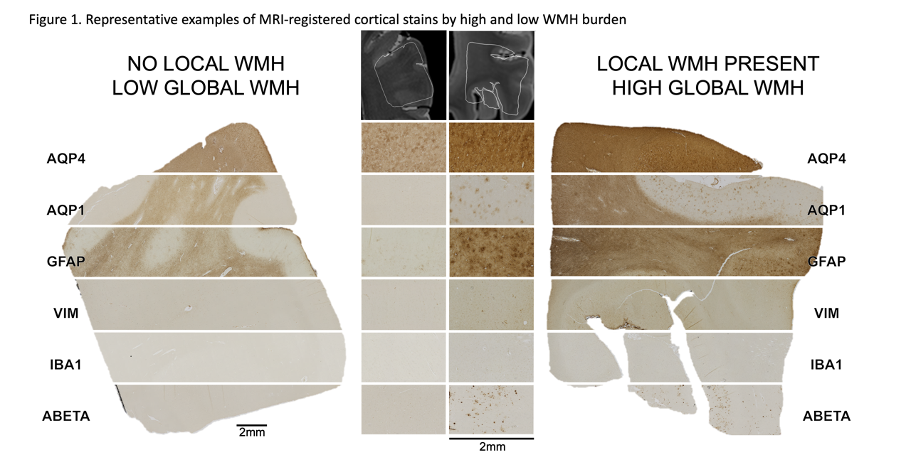
Staining is used on MRI guided samples to characterize pathology. -
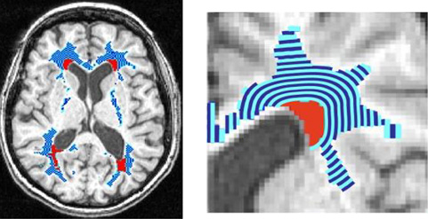
Enlarged perivascular spaces may be related to cerebral small vessel disease and Alzheimer's pathology. We have developed an automated method to detect and quantify the volume of perivascular spaces using multimodal imaging and morphological analysis. By tracking changes in perivascular spaces we will be better able to understand how they relate to cognitive decline and disease processes.
Automated technique to identify enlargement of the perivascular space using intensity thresholding and structural morphology -
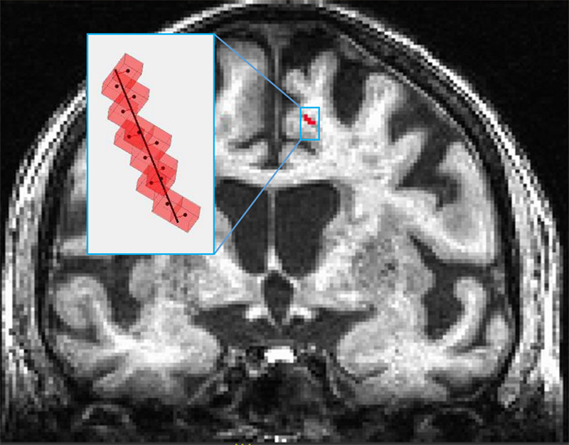
The OADC Neuroimaging Lab has active research examining the relationship between nutrition and brain health by measuring nutrient metabolites in blood and their relationship to MRI makers of brain health, including cerebral blood flow and gray matter density. We are closely involved in clinical trials investigating nutritional interventions, such as fish oil, to maintain brain health with age.
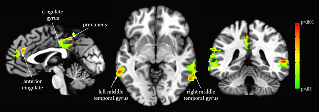
Regions of the brain where grey matter density is related to a metabolite -
OADC neuroimaging lab is using positron emission tomography, utilizing newly developed radioligands, to assess the presence of the amyloid plaques and neurofibrillary tangles associated with Alzheimer's disease, in dementia-free elderly participants.
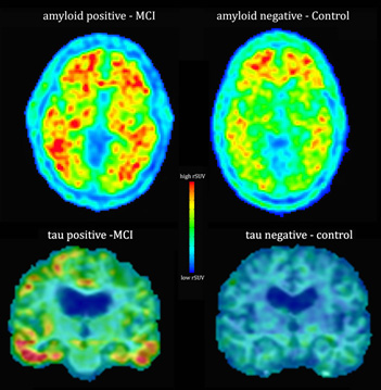
Scan showing amyloid plaques and neurofibrillary tangles associated with Alzheimer's disease