Services and Costs
General information
Please review all information in the Animal Care in the SARIC page of this site prior to use. All protocols performed at the SARIC must be approved by IACUC at OHSU without exception. The laboratory is not currently approved for Biosafety Level >1. Any potential exposures to infectious or toxic agents must be conveyed to SARIC staff. Return of animals to the vivarium after imaging must be approved by the IACUC and the Veterinary staff of the Department of Comparative Medicine (DCM). For animals receiving radionuclides, specific policies have been developed for radiation safety; please contact the SARIC staff to learn about these procedures. Please contact the DCM for any questions regarding return of animals from barrier or quarantine housing. It is recommended that immune compromised animals be studied as early in the scheduled day as possible. Each user is responsible for equipment cleansing.
The hours of operation for the facility are from 9:00 to 5:00 p.m. Monday to Friday. The laboratory is located in room 270 in the access-restricted Vivarium space on the second floor of the Medical Research Building.
Service request
The service request form must be completed for all SARIC activity. For questions, please contact William Packwood 503-494-7803.
Description of services
Use of the ultrasound and biophotonics equipment in the SARIC can be performed on an independent basis or on a supervised basis with technical support provided. Independent use is allowed only after completion of training provided on an ad hoc basis. Supervised use includes technical assistance in system setup, image acquisition, and image analysis. Technical assistance will not be provided for animal handling, administration of anesthesia, or injections. All services for the microPET/SPECT/CT system will be performed on a supervised basis. Investigators must arrange for their own imaging probes. Approval for all radionuclides must be obtained through the Radiation Safety Office at OHSU.
Services and Fees
Biophotonics
Independent use $79/hr; Supervised $157/hr
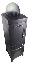
The biophotonics system is a high-efficiency optical imaging camera (IVIS Spectrum, Caliper Life Sciences) originally developed by Xenogen. This system provides non-invasive spatial and longitudinal monitoring of disease progression, cell trafficking and gene expression patterns in living animals.
The IVIS Spectrum at the SARIC contains an optimized set of high efficiency filters and spectral un-mixing algorithms for detecting luminescent or fluorescent reporters across the blue to near infrared wavelength region. The IVIS Spectrum is capable of either trans-illumination (from the bottom) or epi-illumination (from the top) to illuminate in vivo fluorescent sources. Fluorescent 3-D tomography can be performed to determine source localization and concentration using the combination of structured light and trans-illumination fluorescent images.
The instrument is equipped with 12 narrow band excitation filters (35 nm bandwidth) and 24 narrow band emission filters (20 nm bandwidth) that assist in significantly reducing autofluorescence by the spectral scanning of filters and unmixing algorithms. The spectral unmixing tools also allow the researcher to separate signals from multiple fluorescent reporters within the same animal.
Imaging platforms for either rat or mouse imaging have species-appropriate nosecones for inhaled isofluorane anesthesia which is provided through an integrated delivery system. The core facility does not supply fluorophores, luminescent substrates, or injectable anesthetics.
The following are suggested algorithms for luciferin preparation and luciferin detection as well as a fluorescent probe reference that describes the excitation/emission spectral peaks for a broad range of fluorochromes.
Ultrasound
- Independent use $100/hr; Supervised $170/hr
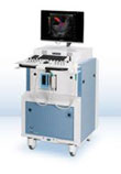
High Frequency Ultrasound imaging is becoming a cornerstone of in vivo small animal imaging because of its ability to provide rapid and real time imaging of tissue function and structure with high resolution. The SARIC houses a dedicated small animal imaging ultrasound system (Vevo 2100, VisualSonics) equipped with phased-array transducers with a frequency range of 18 to 55 MHz. The system is designed for 2-D, 3-D, M-mode, spectral Doppler, color Doppler, and contrast ultrasound; as well as ultrasound-guided microinjection. Common applications include assessment of tissue/organ/tumor size, microvascular perfusion imaging, comprehensive echocardiography, vascular imaging, molecular imaging, and guidance of interventional procedures or microinjection.
The system is equipped with a dedicated inhaled anesthesia system with noscones for inhaled isoflurane for mouse or rat imaging. There are mouse and rat integrated physiology platforms that are heated and capable of monitoring ECG, respiratory rate, and body temperature. The railed imaging gantry is capable of x-, y-, z-adjustments in the imaging plane and co-registration of ultrasound imaging probes with microinjector systems. The system also contains an integrated motor system for elevational plane adjustments that provides 3-D reconstruction.
The imaging system has a broad frequency range with low frequencies for better penetration in rat models and contrast sensitivity, and high frequency range for better spatial resolution (30 microns spatial resolution). The system is equipped with contrast detection software and online Doppler and 2-D analysis packages. Pulsed-wave and power Doppler technologies are available for vascular imaging.
MicroPET/SPECT/CT
- $425/hr PET and/or SPECT Protocols - Supervised use only
- $170/hr CT only Protocols - Supervised use only
The SARIC houses a state-of-the-art integrated small animal imaging platform for near simultaneous and co-registered radionuclide SPECT, PET and CT imaging (Inveon, Seimens). The Inveon (Siemens) imaging system is capable of true trimodal imaging guided by computer positions systems located within the gantry. The system has an integrated system for inhaled anesthesia and maintenance of isothermia. There is an onsite radionuclide hot lab for dosimetry, cold kit preparation, and storage. Typical applications include high-resolution CT anatomic imaging, cell tracking, perfusion imaging, molecular imaging, cell proliferation, enzyme activity, pharmacodynamics, gene profiling, and metabolic imaging.
The CT imaging system is a single integrated gantry with a 14 bit x-ray imaging detector with 4,096 X 4,096 pixels (10 X 10 cm FOV) for imaging not only small but large rodents. The system has variable focus x-ray source for spatial resolution down to 20 microns. There is also a high-performance work station with real-time and scalable reconstruction with 512 X 512 X 768 voxel volumes.
For SPECT imaging there is a 2-headed detector (15 X 15 cm) system for whole animal or high-magnification studies. Gamma ray detection range is from 30 to 300 keV. The system has a general purpose, mouse pinhole, and rat pinhole collimator.
For PET imaging, there is a high performance lutetium oxyorthosilicate (LSO) crystal detector with absolute sensitivity of 10% at the center of the FOV. The detector pixel spacing (1.59 X 1.59 mm) provides a 1.4 mm full width at half-maximum spatial resolution using a traditional filtered back projection algorithm. An axial FOV up to 30 cm is possible.
The research workplace software provides imaging review, image fusion (including 3-D fusion images of CT, SPECT and PET), and analysis packages. Radionuclide data can be processed with either filtered back projection or alternatively with iterative reconstruction (ordered-subset expectation maximization [OSEM]) processing for better signal-to-noise or shorter acquisition times. CT attenuation correction algorithms are used.
Although the director of the SARIC maintains a nuclear license for the facility, approval of all protocols employing radionuclides must be obtained through the Radiation Safety Office at OHSU. All investigators must be familiar with SARIC post-imaging handling procedures for animals receiving radionuclides.