About us
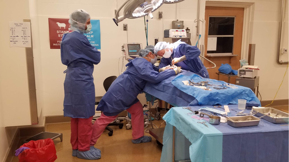
Sonnet Jonker's research focuses on heart growth and function before and around the time of birth because these months are critical for establishing life-long cardiovascular health.
Before birth, working cardiac myocytes proliferate abundantly, the coronary capillary tree expands, connective tissues are deposited in the heart and blood vessels throughout the body. The fetus is exquisitely responsive to its environment, though, and has the capacity to respond by altering growth and maturation in mere days. Adaptations help the fetus to meet a challenge, but may have long-term consequences. We are interested in how the fetus responds to adverse challenges, such as placental insufficiency, anemia, and congenital cardiac malformations, and whether we can improve their life-long health by stimulating beneficial adaptations in the perinatal period.
Check out our fantastic team, as well as our publications and on-going projects.
We're moving!
We're excited to announce that the lab will be joining the Lawrence D. Longo MD Center for Perinatal Biology! Sonnet, Sam, Sarah and Neeka will be relocating to Loma Linda University on July 1, 2025. We will be continuing all our research projects to understand fetal and neonatal cardiovascular development with new colleagues. While sad to be leaving beautiful Portland, we are looking forward to new adventures and opportunity for growth.
Funded by
NIH / National Heart, Lung, and Blood Institute
Collins Medical Trust
Additional Ventures
Equity
Sonnet Jonker, Sam Louey, and the members of the Jonker Lab are committed to making careers in biomedical research accessible to everyone, no matter their race, culture, faith, sexual orientation, or gender identity and expression, by actively supporting equitable access to training, collaboration, networking, appointment, promotion, and a positive work environment.
DOHaD Summer Course
The DOHaD Summer Course is designed for graduate students, post-doctoral fellows and early-career faculty who are interested in the long-term consequences of developmental programming. The course introduces participants to different perspectives within the field of DOHaD thru a series of lectures by nationally recognized researchers.
What's new
- Joseph presents at the American Physiology Summit
- Eric presents Pediatric Grand Rounds
- Eric and Sonnet present at Society for Reproductive Investigation
- Published: KNDy Neurons and the control of the gonadotropic axis in the midgestation fetal sheep
- Additional Ventures Expansion Award to study flow distribution