Brain and Spinal Tumors
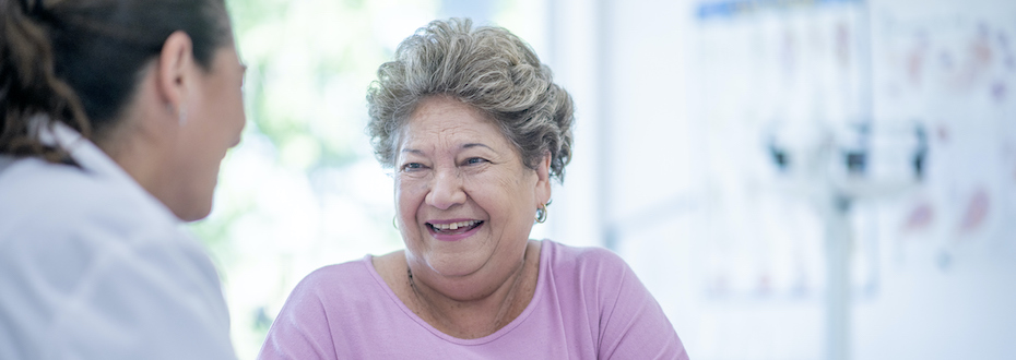
The OHSU Brain Institute offers expert, compassionate care for brain and spinal cord tumors. We use advanced techniques to treat tumors while protecting healthy tissue.
At the Brain Institute, you’ll find:
- Team-based care, with many specialists working together.
- Neurosurgeons who focus only on brain and spinal cord tumors. They are experts in complex procedures.
- Advanced technology, including MRI used during surgery.
- Innovative methods, including one we pioneered to get medications past the blood-brain barrier.
- A commitment to research, turning our discoveries into better patient care.
Learn more
Our excellence
Knight Cancer Institute partnership: Our doctors are also part of the OHSU Knight Cancer Institute.
Precise diagnosis: Our in-house lab helps us make a fast and accurate diagnosis. We can analyze a tumor’s genetic makeup to choose the most effective treatment.
Advanced treatments: We perform innovative surgeries, including awake craniotomies, to remove tumors while protecting brain function. You’ll also have access to new ways of delivering chemotherapy and relieving symptoms.
Patient-centered care: We get to know you and your goals so we can plan the treatment path that is right for you.
Team-based care
Our team of specialists gets you the care you need. Your team may include neuro-oncologists, neurosurgeons, neuroradiologists, radiation oncologists and neuropathologists.
Tumor board: Specialists meet each week to review patient cases one at a time. They combine their expertise so you can benefit from the full team’s knowledge.
Prompt results: We get your test results quickly so you won’t have to wait for answers. Some results are ready the same day.
Leading-edge treatments
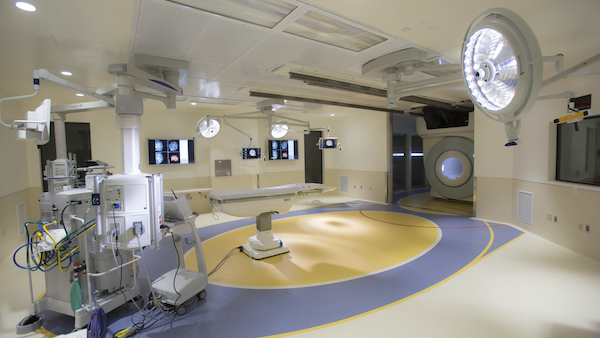
Intraoperative MRI: This technology lets our surgeons remove tumors with remarkable precision. Our iMRI scanner, located in Doernbecher Children’s Hospital, is available for adult surgeries.
PET-MRI: This technology, not widely available elsewhere, lets your care team track tumor growth and follow the path of medications. This helps us plan your treatment.
Brain mapping: Our team’s careful planning for brain surgery includes mapping your brain. We use imaging to find the tumor before surgery. During surgery, we use a small electrical probe to find the areas in your brain that control speech and movement. We avoid those areas as we remove your tumor.
Blood-brain barrier disruption chemotherapy: The blood-brain barrier prevents chemotherapy from reaching brain tumors. Our researchers found a way to open the barrier to let the medication in. The Brain Institute is a world leader in this treatment.
Targeted radiation therapy: We use one of the most advanced radiation therapy systems available. We point the radiation beam directly at the tumor so we can use a high dose while avoiding healthy brain tissue.
High-resolution imaging: We use a contrast agent (dye) that we studied right here at the Brain Institute. It lets us see your brain in great detail. We can see if treatment is working and plan the next steps.
Minimally invasive procedures: We provide MRI-guided laser ablation, a less invasive option to treat certain types of brain tumors.
Clinical trials and research
If you are eligible for any clinical trials, we’ll discuss them with you so you understand all your options. We are studying:
Chemoprotection: We are finding ways to avoid some side effects of chemotherapy. One technique may help protect hearing as we treat a brain tumor.
Targeted chemotherapy: This approach delivers the medication only to the brain instead of through the whole body.
Immunotherapy: We can use the power of your immune system to destroy tumors. Some of our studies:
- Put medication into the tumor to activate the immune system.
- Combine radiation therapy with medications that help your immune system find and attack cancer cells.
- Use advanced imaging to quickly see if a treatment is working. If not, we can stop right away and try something else.
Palliative care
You have access to palliative care at any point during your treatment. We can help you make hard health care decisions, ease your pain and relieve stress. We want you to be as comfortable as possible.
For patients
- Referral: To become a patient, please ask your doctor for a referral.
- Questions: For questions or follow-up appointments, call 503-494-5626.
Location
Parking is free for patients and their visitors.
OHSU Neuro-Oncology Clinic, Marquam Hill
3270 SW Pavilion Loop, 2nd floor
Portland, OR 97239
Map and directions
Refer a patient
- Refer your patient to OHSU.
- Call 503-494-4567 to seek provider-to-provider advice.