Primate Multimodality Imaging Center
About us
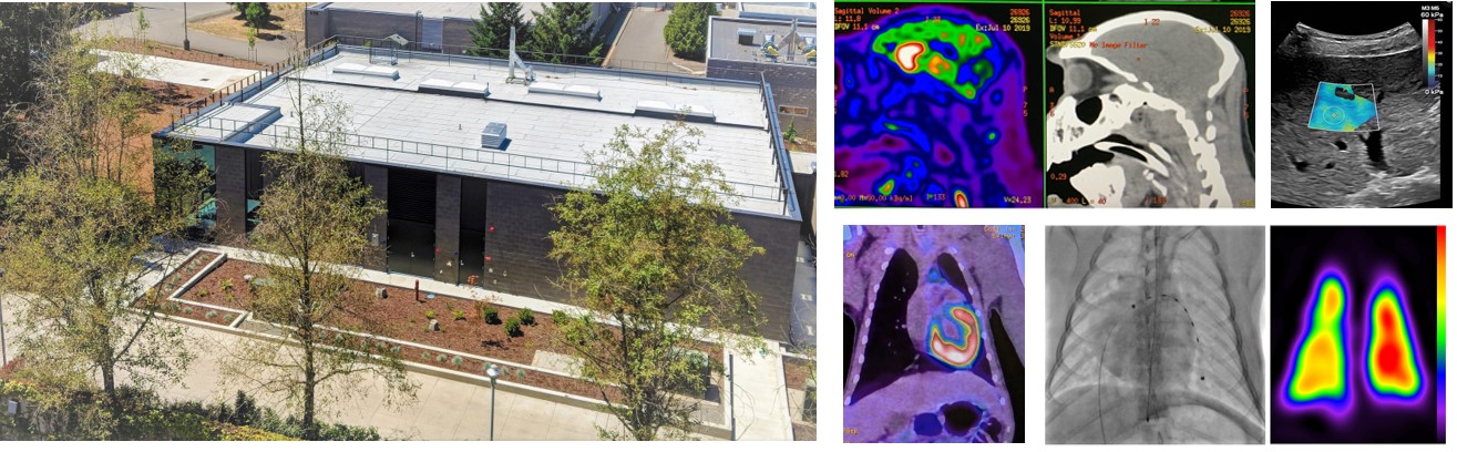
The Primate Multimodality Imaging Center (PMIC) is a comprehensive imaging facility dedicated to state-of-the-art non-invasive imaging. The center is located in a building designed specifically for in vivo imaging of nonhuman primates with biplane angiography, PET/CT, SPECT/CT, ultrasound, and DEXA, together with support laboratories (wet lab, radionuclide lab). The center is directly adjacent to the ONPRC Magnetic Resonance Imaging (MRI) center, with direct hallway transport to the Animal Services Building (ASB) that houses non-human primates and advanced surgical facilities.
The PMIC is designed to provide not only imaging equipment and image analysis software, but also expert guidance with respect to selection of appropriate imaging modalities, image acquisition and analysis protocols, contrast agent selection and development, and coordination with the OHSU Center for Radiochemistry Research for radionuclide probes. All services involving radionuclide imaging (PET, SPECT), angiography, or CT require a Facilitation Meeting with imaging experts prior to initiating studies in order to establish image acquisition protocols, animal welfare, and coordination with CRR or other resources. These meetings can be scheduled prior to final IACUC approval.
Contact us
PMIC Interim Director: Charles Roberts, Ph.D., robertsc@ohsu.edu
PMIC Manager: John Templon, CNMT, R.T.(CT), templonj@ohsu.edu
office: 503-346-5301
PMIC Staff: Jim Hodovan, Bill Packwood
Facilitation Meeting Scheduling: Mary Postiff, postiff@ohsu.edu
PMIC oversight committee
- John Templon, CNMT, R.T.(CT)
- Lauren "Drew" Martin, DVM
- John Kaufmann, MD
- Alison Weiss, Ph.D., Chair
PMIC Services
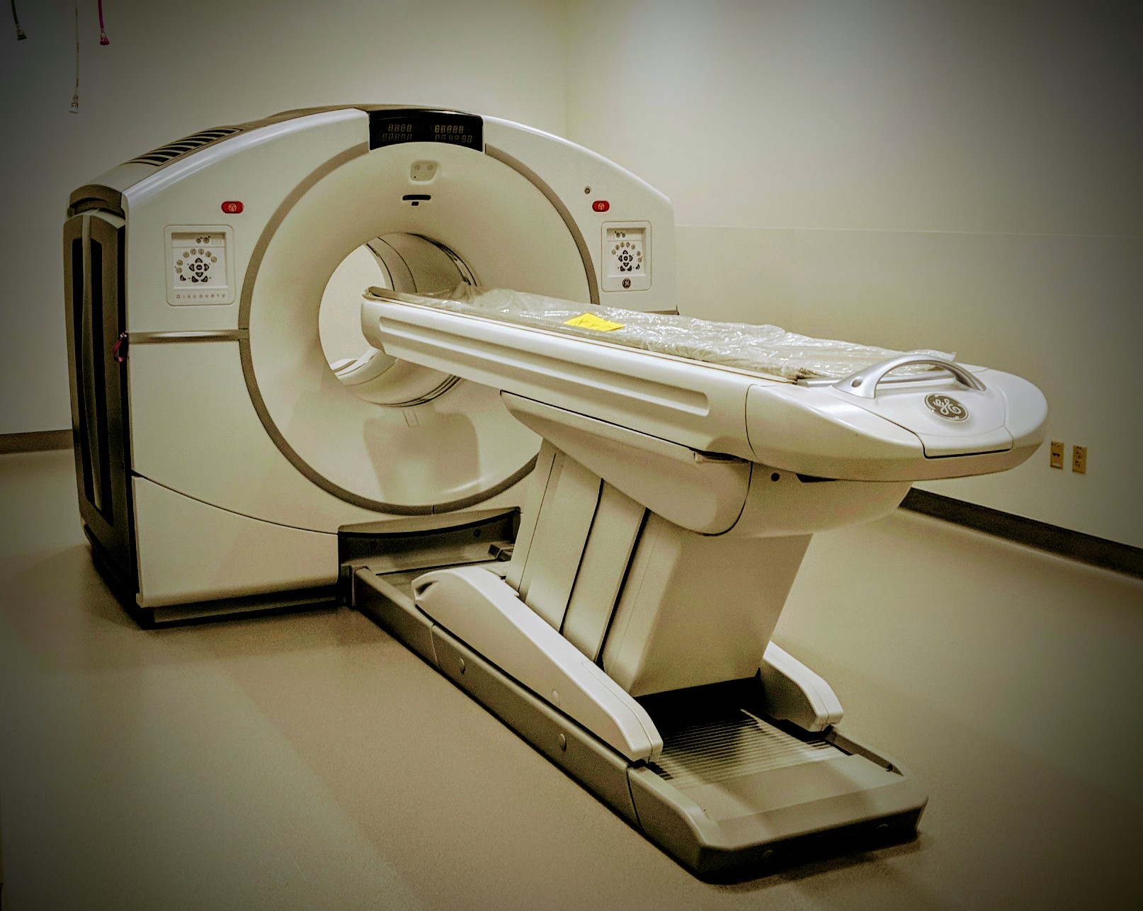
GE Discovery MI 710 PET/CT
Hybrid Imaging System
The system consists of an integrated gantry containing 24 PET detector rings incorporating next-generation solid state detectors and an Optima CT 660 with a 64-slice detector. The axial and trans-axial fields-of-view (FOV) on the system are 15.7 and 70 cm, respectively. There are PET imaging planes with user-defined slice overlap (1 to 23).
This system includes prospective reconstruction with VUE Point HD which provides 3D iterative reconstruction and 3D scatter correction. WideView technology allows PET reconstructed FOV coverage of 70 cm diameter with CT attenuation correction. The Q.Clear technology on the system is a full convergence iterative reconstruction algorithm that maximizes SNR, while the SharpIR incorporates information from the PET detector point-of-spread function in order to improve spatial resolution when working with nonhuman primates. The ImageWorksTM module provides access to advance image processing features, including CT perfusion and bone analysis.
The system can also be operated in CT stand-alone mode (512x512 reconstruction matrix) and includes 5-beat cardiac mode (44ms temporal resolution).
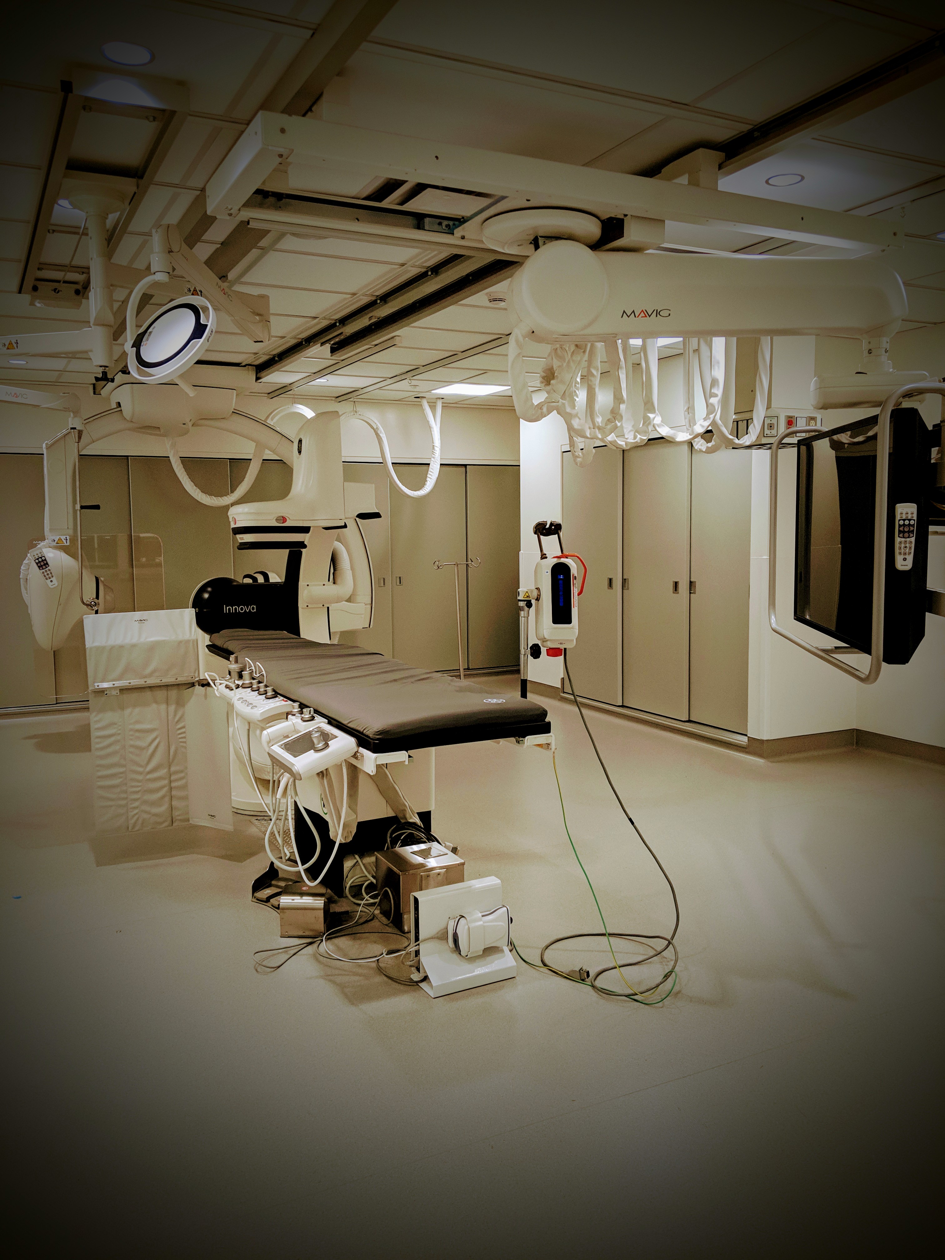
PMIC Angiography Suite with the GE Innova IGS 620
Bi-plane Angiography
The system is fully equipped for cardiac, vascular and interventional X-ray imaging and consists of floor- and ceiling-mounted C-arms with 20.5 x 20.5 cm Innova solid state digital detectors. This system has a 3-axis iso-centric positioner gantry design for the frontal and lateral positioners for optimizing positioning flexibility. A state-of-the-art table is designed for low adsorption and scatter with full angulation capacity and complete tableside status control. The X-ray tubes are 160A metal and fluoroscopic power is 3200 W (continuous) and 4500 W (peak, 10 min). The system has a silicone photodiode array (1024 x 1024) with a FOV of 12-20 cm. The system includes 2 pairs of collimation blades and contour filters blades with free rotation. There is a wide dynamic range option on the system (“DRM”) with 14 bit acquisition. Fluoroscopy can be performed without DSA with a frame rate of 7.5-30 fps for single plane and 3.75-25 fps in biplane. Advanced features include stenosis quantification, runoff analysis, blended roadmaps for superimposed vascular maps, and subtracted 3D image visualization.
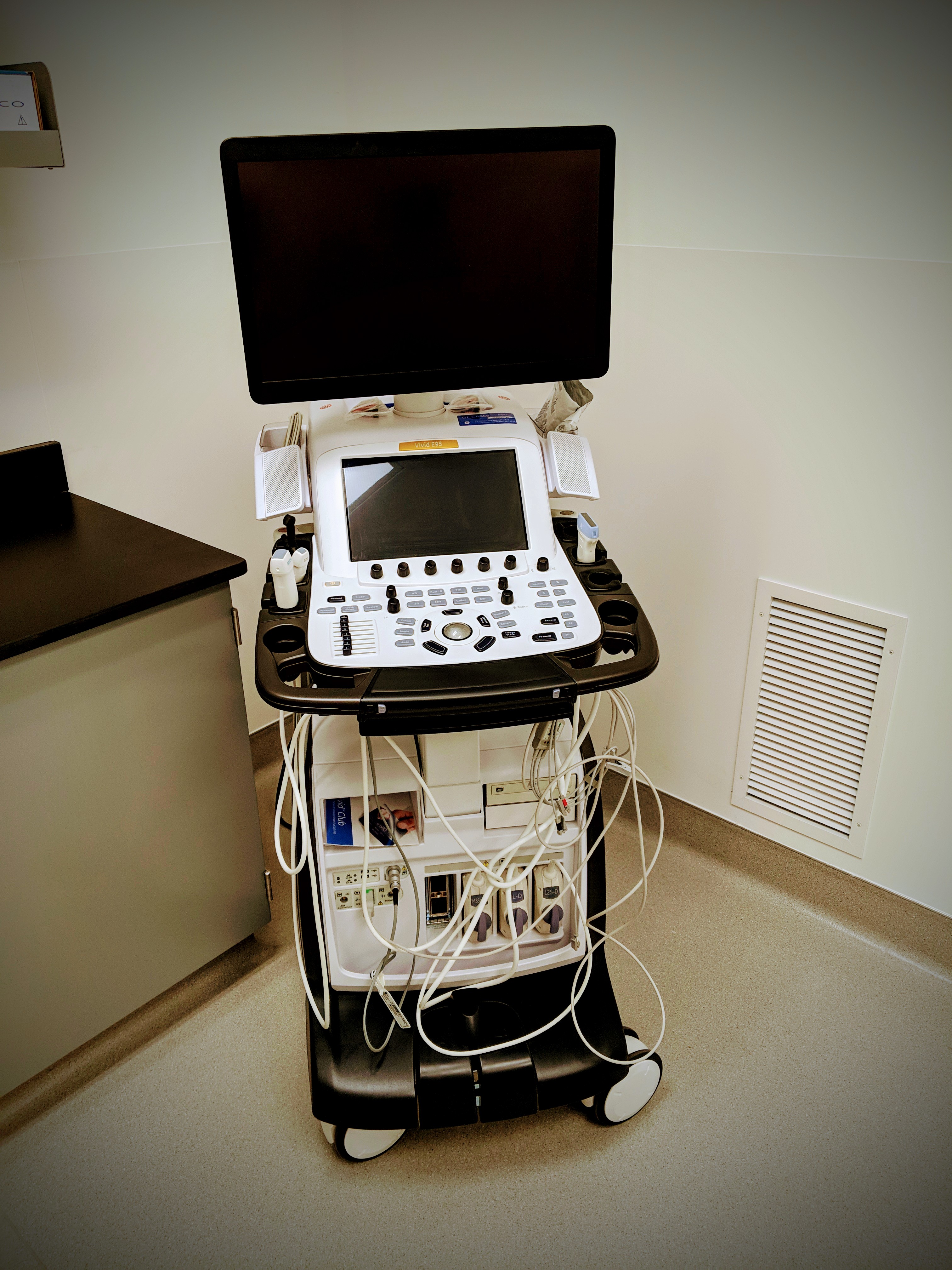
- GE Vivid E95
- GE Logiq E9
- Siemens Accuson Sequoia 512
- Philips Sonos 5500
There is a wide array of ultrasound systems in the PMIC, which allows flexibility according to the strengths/features of each system and specific imaging needs. These systems allow the selection of the appropriate system based on considerations such as spatial resolution (frequency), vascular imaging, depth/power, 3-D imaging, strain analysis, perfusion imaging (high or low resolution), advanced tissue analysis (elastography, shear-wave elastography, integrated backscatter, full RF output), transducer needs (linear-array, curvilinear-array, phased-array, Pedoff, vaginal, TEE), stress packages, signal processing capability (harmonic imaging, power-harmonic Doppler, phase-inversion, power-modulation, tissue Doppler imaging, speckle strain analysis, maximum intensity projection analysis, etc.).
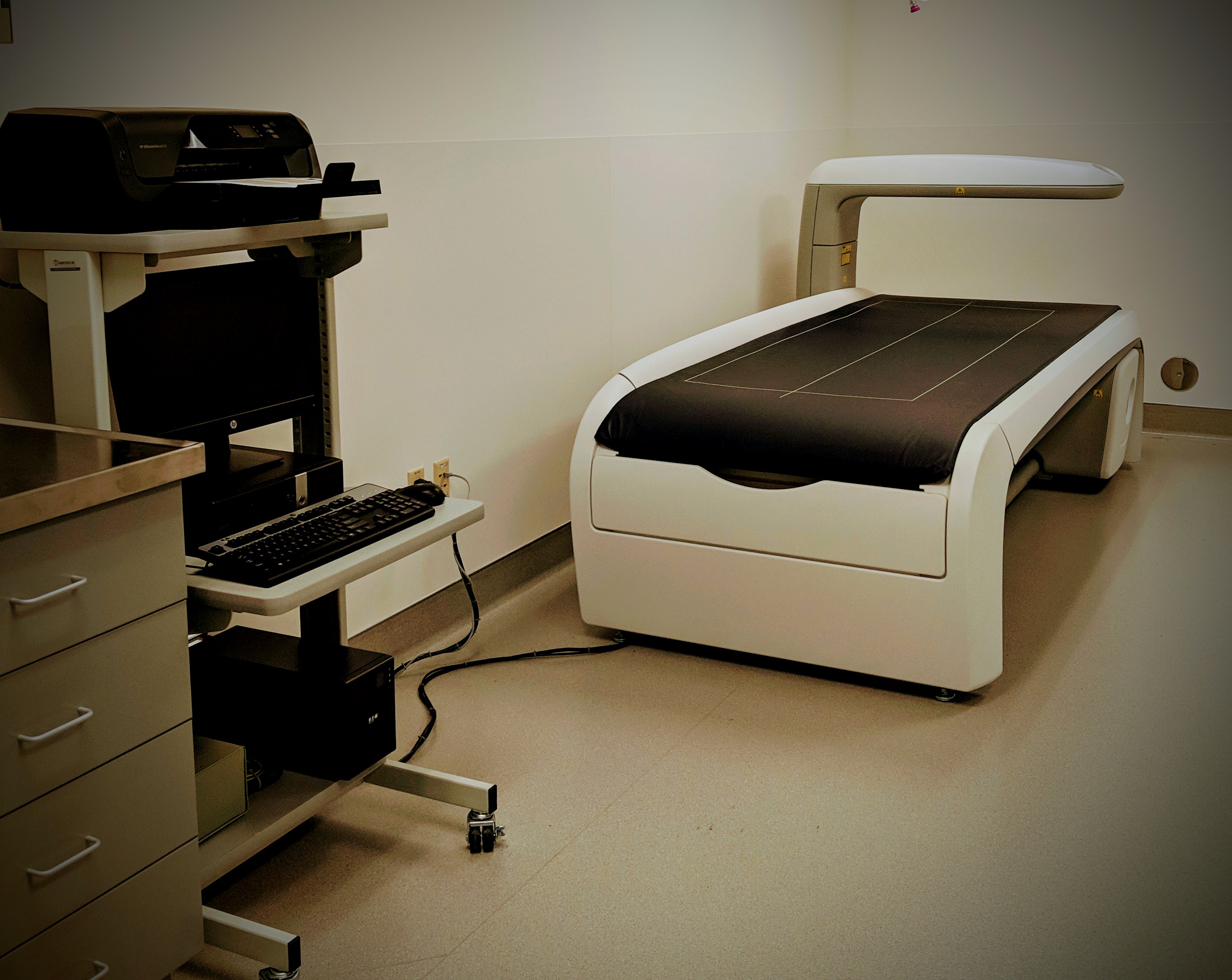
GE Lunar iDXA Advance system
A research-grade direct-digital fan-beam dual-energy x-ray imaging system. This system is designed for a variety of advanced applications, including analysis algorithms for bone densitometry, body composition, and visceral fat. This system has specific pediatric imaging capabilities which are critical for studying non-human primates.
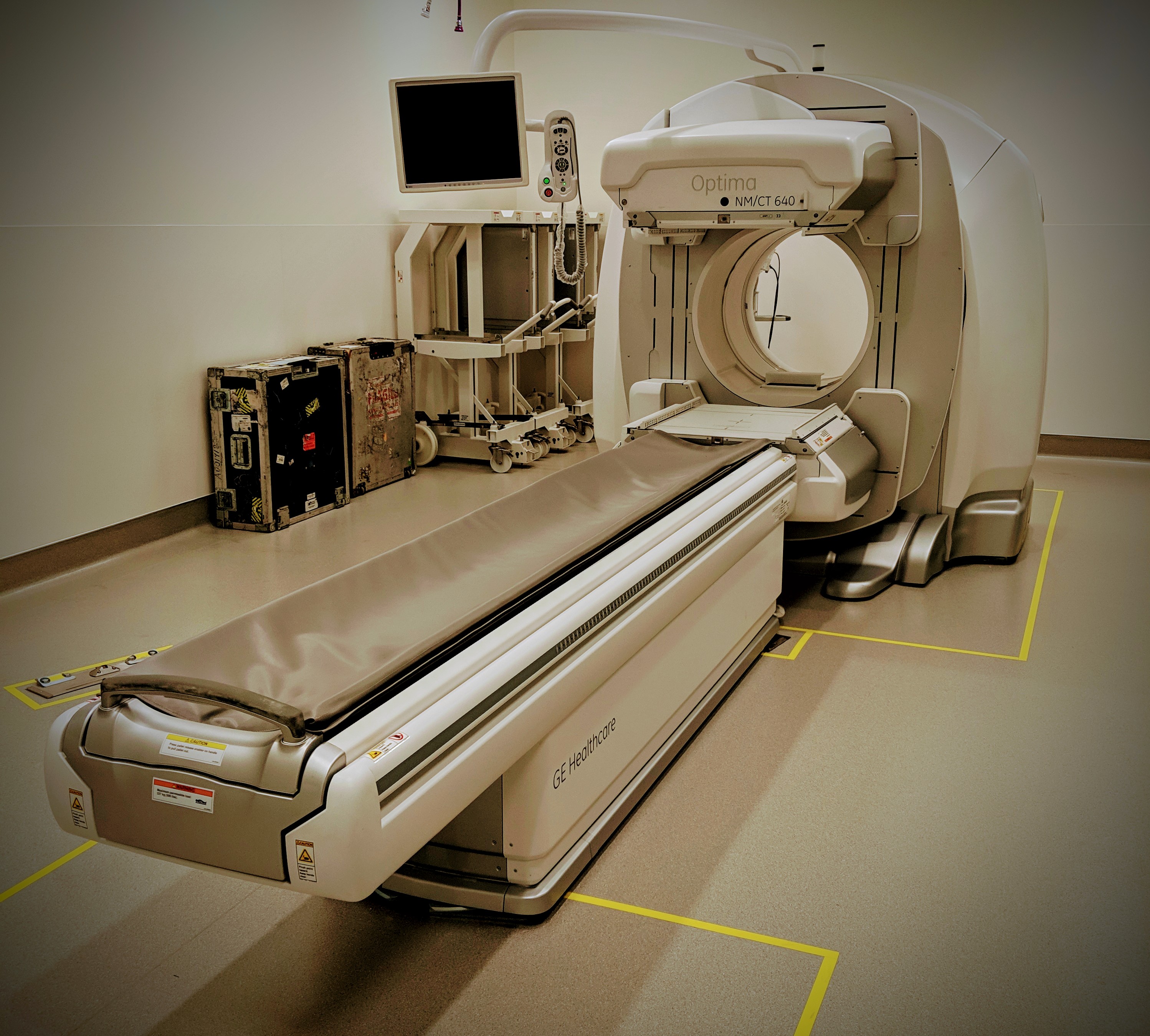
GE Optima Hybrid 640
Single-Photon Emission Computed Tomography (SPECT)
An all-purpose, dual detector, free-geometry nuclear imaging system. The gamma detectors on the system are digital Elite NXT technology on a slim 70 NM gantry integrated with a high-performance CT 640 system (8-slice).
This system enables acquisition of SPECT, whole-body planar, and gated SPECT/planar studies. Detector geometry (e.g. 90°, 180°, and custom angles) and radial motion are of flexible design. There is a real-time infrared-based automatic body contouring designed to enhance scanning efficiency and detector distance. Key features of the NM gantry are a bore size of 70 cm, maximal rotational speed of 0.033 to 3.0 rpm, and rotation range of 540°. The Elixt NXT detectors are composed of 3/8” NaI crystal (9.5 mm) with 59 circular photomultiplier tubes (76x38 mm), a 30 MHz sampling rate, and an energy range of 40-620keV. The Optima CT 640 CT sub-system includes 0.625 mm FWHM helical reconstruction.
To enable flexibility of use, the system includes LEHR collimators, MEGP collimators, and a Fan-beam collimator. Beam tracking technology allows real-time X-ray. Full analytic and post-processing capabilities are included with the system including NM-CT registration, motion correction, CT Attenuation Correction (CTAC), 3D display, gated analysis, and image co-registration with other technologies.