Orthodontics at OHSU
The orthodontics program houses a modern clinical faculty with a diverse faculty who have a combined wealth of experience. Patients of all ages receive expert and compassionate care. School of Dentistry students take part in rigorous advanced education to prepare them for a lifetime of learning and service.
-
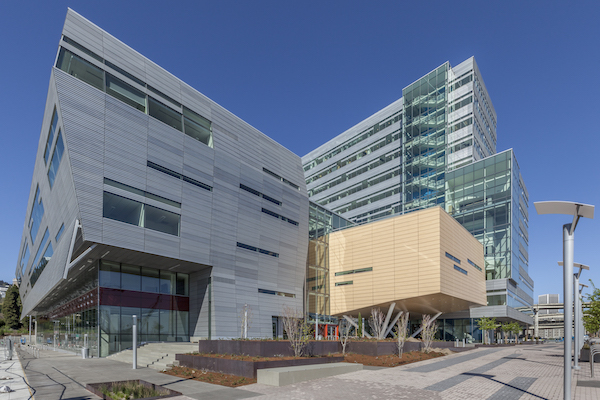
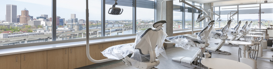
The Oregon Health & Science University’s Division of Orthodontics and its specialty clinic are housed in the Robertson Life Sciences Building on the South Waterfront in Portland, Oregon. Patients benefit from evidence-based, customized treatment from our dedicated and diverse faculty and staff.
Talented pre-doctoral education and post-doctoral students come from all reaches of the globe join our graduate program because of its vibrant culture. Continuous course updates and cutting-edge research results in superior clinical and didactic education, discovery and a commitment to improve access and dental health for our patients.
Activities of the division are strengthened through support by the School of Dentistry Alumni Leadership Council and orthodontists in the region.
-
Orthodontics leadership
Laura R. Iwasaki, D.D.S., M.Sc., Ph.D., professor-provisional, department chair
Jeffrey C. Nickel, D.M.D., M.Sc., Ph.D., professor-provisional, director of advanced education in orthodontics program
Howard Freedman, D.D.S., assistant professor, director of orthodontic clinics
-
Master of Science
The master of science program in orthodontics qualifies candidates for specialty practice and/or academic careers and for examination for certification by the American Board of Orthodontics. It is a 30-month continuous program (10 academic quarters) and consists of course work in the basic sciences, laboratory, theory, clinic and research in related fields. Learn about the Student Learning Outcomes of the orthodontics master's program.
Selection Process
More information on applying to our Advanced Education programs
Faculty of the Department of Orthodontics will review completed applications received in the Office of Admissions by the deadline specified by PASS. From the submitted materials, applicants will be selected for invitations to interview. The selection criteria for interviews are multifaceted, taking into account all the application materials. Interviews typically take place in early November. Those selected will be sent invitations, usually by early October. Following interviews, applicants will be rank ordered by the department and submitted to the postdoctoral dental matching program.
Course Offerings
View a PDF of our graduate program course offerings.
Additional Information
Dr. Jeffrey Nickel
Graduate Program Director
Department of Orthodontics
OHSU School of Dentistry
2730 S.W. Moody Avenue, SD-Ortho
Portland, Oregon 97201-5042Direct questions about the application process to:
Office of Admissions and Student Affairs
OHSU School of Dentistry
2730 S.W. Moody Avenue
Portland, Oregon 97201-5042
Support Orthodontics
Donate to support the Orthodontics Division and resident education.
Connect with us
Clinic Line: 503-494-8867
2730 S. Moody Avenue
Portland, Oregon 97201
Read OHSU Dental Clinic’s patient appointment instructions before arriving for your scheduled appointment.
Contact Us
OHSU School of Dentistry
Department of Orthodontics
2730 S. Moody Avenue
Portland, OR 97201-5042
Graduate Program Director
Jeffrey Nickel, D.M.D, M.Sc., Ph.D.
Email Dr. Nickel