Endodontics at OHSU

The OHSU School of Dentistry Advanced Education Program in Endodontics trains dentists to become competent entry-level endodontists who are committed to preserving the natural dentition of a diverse patient population. We aim to excel in curricular innovation, achieve clinical excellence using contemporary techniques and be recognized as a scholarly program both nationally and internationally.
Table of Contents
About the Endodontics Department
Endodontics at OHSU School of Dentistry involves root canal treatment for patients by undergraduate dental students and by the graduate residency program. Cases handled by undergraduate dental student involve root canal treatment only under the guidance of a specialist in endodontics in the undergraduate clinic. The graduate residency program provides treatment for patients with more difficult cases. Root canal therapy, retreatment of existing cases and surgical procedures, if necessary, are completed in the graduate program clinic.
Purpose
Our purpose is to educate endodontic oral health care specialists in the provision of comprehensive, evidence-based endodontic care, and who undertake lifelong learning, ethical clinical practice, community service and leadership.
Mission
Our mission is to engage in evidence-based didactic and clinical education, contemporary research, compassionate interdisciplinary patient care and healthcare advocacy for the community and the profession.
The OHSU Division of Endodontology comprises a diverse faculty group dedicated to teaching and mentoring dental students and residents in providing compassionate endodontic care to relieve pain and suffering, while retaining the natural dentition. Our graduate students and faculty work collaboratively with other disciplines to achieve better patient outcomes and foster intellectual curiosity.
Our Team
- Adam Lloyd, B.D.S, M.S.
Professor provisional, division chair of endodontology
Program director in the Advanced Education Program in Endodontics
lloydad@ohsu.edu - Faculty
- Residents
The Endodontics Graduate Program
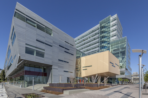
Located on the 11th floor of the Robertson Life Sciences Building at the South Waterfront, the graduate endodontic clinic provides state of the art treatment for patients and education for students and residents. Usage of modern amenities includes dental operating microscopes, a dedicated CBCT unit, ASI carts, individualized operatories and a dedicated surgical operatory. Our team approach fosters inquisitive thinking and referrals from inside the School of Dentistry and the community.
Interdisciplinary treatment planning occurs in conferences with periodontology, pediatric dentistry, GPR and orthodontic graduate students. A hallmark of our program is treating the medically complex patient, interpreting objective data and special testing, with communication of the findings and treatment to the referring office. A typical graduate will complete 175 non-surgical root canal treatments, 35 retreatments and 15 surgical cases. Exposure to vital pulp therapy in collaboration with pediatric dentistry residents, and IV sedation with the GPR residents is expected. Surgical cases are performed by GS2 residents.
Endodontology faculty will review completed applications received in the Office of Admissions by the deadline specified by PASS. From the submitted materials, applicants will be selected for invitations to interview. Only individuals who have completed their dental degree program at a CODA accredited school, before the PASS deadline, will be considered for an interview. Selection criteria for interviews are multifaceted, taking into account all the application materials.
Learn about the student learning outcomes of the Advanced Education Program in Endodontics.
Read about the graduate program course offerings.
Applicant guidelines
Our certificate program complies with standards established by the American Dental Association’s Commission on Dental Accreditation, or CODA, and qualifies residents for examination by the American Board of Endodontics. Continuous full-time attendance is a requisite for the 24 month course of instruction. Three graduate students are accepted each year. Details regarding application to the program can be found through the PASS Portal. The PASS portal typically opens mid-May, with our deadline July 1. The applications are reviewed on a rolling basis. The program’s start date is July 1 of the anticipated year of matriculation. The certificate program lasts (a minimum of) two years from the date of entry, which is July 1. Only individuals who have completed their dental degree program at a CODA accredited school, prior to anticipated matriculation at OHSU, will be considered for an interview. Selection criteria for interviews are multifaceted and consider all the application materials, with preference given to candidates with experience.
For further information about the program, please visit the prospective student page.
Awards
American Association of Endodontics Award
The American Association of Endodontics Award, or AAE, is presented to a fourth-year dental student based on a) the summation of all grades earned in endodontics, b) total number of endodontic points earned in the clinic, and c) evaluation of the candidate’s clinical knowledge of endodontics demonstrated in a case presentation contest delivered to endodontic faculty. Candidates receive an AAE certificate and subscription to the Journal of Endodontics. All fourth-year students are eligible.
Sorum-Xia Award in Endodontics
Fourth-year dental students who show exceptional abilities in endodontics are eligible for the Sorum-Xia Award. The students must participate in a case presentation contest delivered to the endodontic faculty. Candidates receive a financial award and certificate, and their name is inscribed on a plaque on display in the School of Dentistry. The award is in honor of Larry Sorum ’78, an endodontist who served in the Air Force and died of cancer at an early age. Tian Xia, D.D.S., M.S., ’02, a School of Dentistry Endodontic Graduate program alumnus, is a donor of the award.
Make a Gift
Leslie A. Morgan Endodontic Endowment Income Fund
The proceeds from the endowment provide essential support for the endodontics residency program. Funds allow endodontic residents to attend the annual American Association of Endodontics conference and board review course meetings, and to support research projects, honorariums, consultants and speaker expenses, as well as special materials and equipment.
Thank you for your generous support. Each year, 5% of the Morgan Endodontic Endowment Income Fund is available to advance educational and professional development of our endodontic residents.
Make a Gift Today
Contribute to the Morgan Endodontic Endowment Income Fund.
About Les Morgan
Les Morgan, D.M.D. ’79, dedicated 14 years to combining his passion for academics and clinical practice at the OHSU School of Dentistry. As an assistant professor in the Department of Endodontology, was deeply committed to the graduate program—mentoring student researchers, delivering lectures and contributing to the clinic.
An accomplished researcher, he authored numerous scientific publications. His early interest in research began during his dental education, when he worked summers as a research assistant at OHSU. Following graduation, he served as a general practice resident with the U.S. Army at Fort Riley in Kansas, before pursuing advanced endodontic training at the University of Texas at San Antonio.
Morgan passed away in 1998, leaving a legacy on the OHSU community through his dedication to education, research and patient care.
Connect with us
Clinic Line: 503-494-8867
2730 S. Moody Avenue
Portland, Oregon 97201
Maps and directions
Read OHSU Dental Clinic’s patient appointment instructions before arriving for your scheduled appointment.
More questions about your appointment? Contact us by email.
Contact us
OHSU School of Dentistry
Department of Endodontology
2730 S. Moody Avenue
Portland, Oregon 97201-5042
Support an endodontics scholarship
Contribute to the Gordon Marshall Endowed Scholarship in Endodontics.
Support excellence in endodontics
Contribute to the Ardon Overby Endowment for Excellence in Endodontics.
Support advanced education in endodontics
Contribute to the Morgan Endodontic Endowment Income Fund.