About
The Advanced Light Microscopy Core at the Jungers Center was established in 2009 through collaborative efforts of the University Shared Resources Program and the Departments of Neurology and Molecular Microbiology and Immunology, the Jungers Center for Neurosciences Research, and the Oregon Hearing Research Center. Technical expertise and instrumentation was pulled into a shared resource to serve OHSU researchers in need of fluorescence microscopy.
Meet the Staff
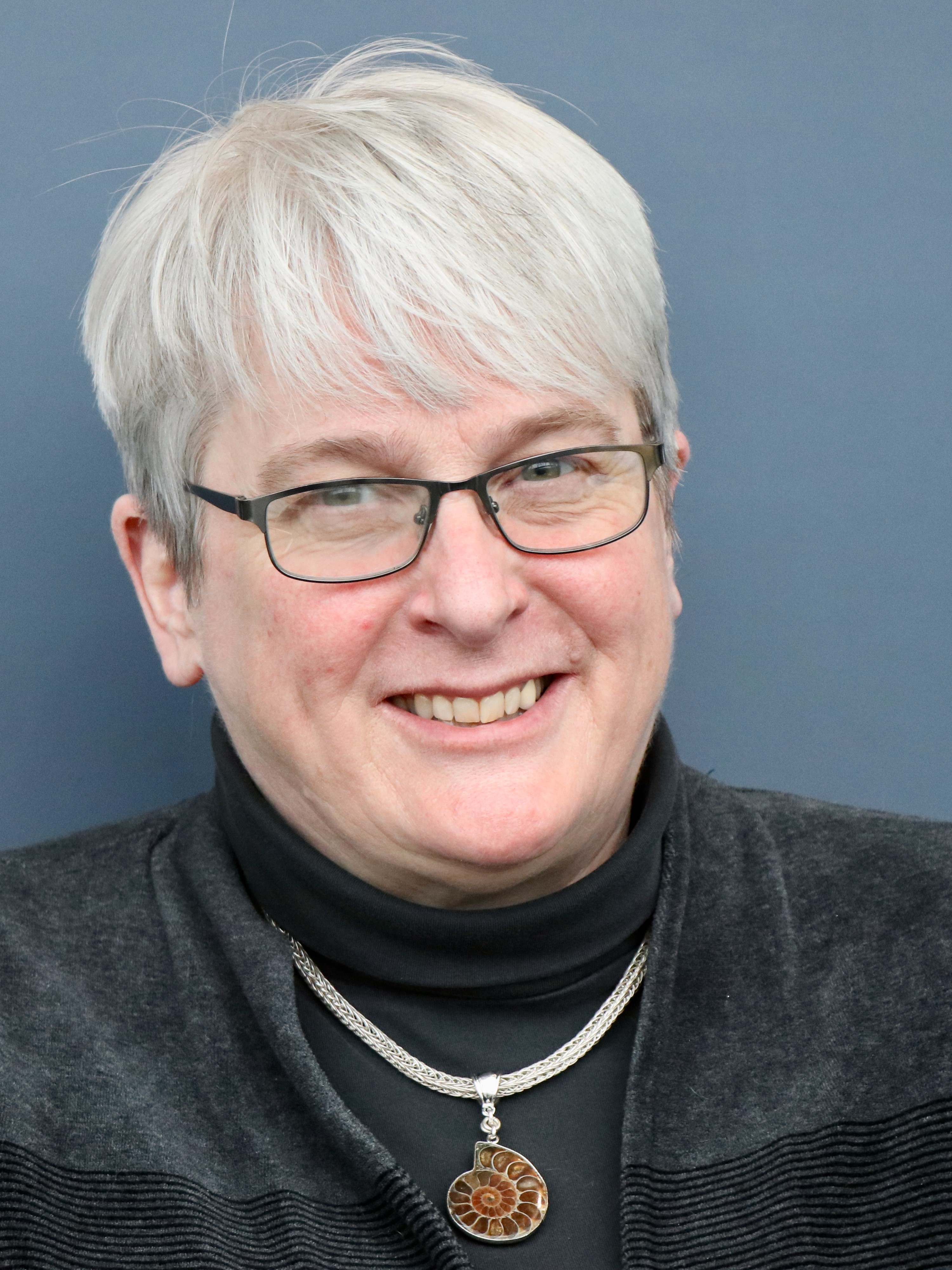
Stefanie Kaech-Petrie: Director

Stefanie puts 100% effort into developing and running programs to enhance advanced light microscopy at OHSU. She has over three decades experience with advanced instrumentation for light microscopy and was among the first neuroscientists to take up the use of GFP technology. Her past research includes visualizing microtubule- and actin-based cytoskeletal rearrangements and membrane protein trafficking in neurons. Contact Stefanie
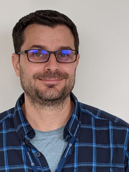
Brian Jenkins: Senior Core Scientist

Brian’s imaging experience spans 10+ years in the neuroscience field using several microscope modalities with a focus in live-cell imaging. Brian’s passion for microscopy began as an undergraduate in San Diego, further developed during graduate school in Oregon, and sharpened as a postdoc in Wisconsin. His main interests reside in how the neuronal cytoskeleton influences protein trafficking and neuronal morphology. In the core, he enjoys assisting researchers image their samples in a meaningful way while teaching the basics of light microscopy. Brian can usually be found at Marquam Hill with a cup of decaf in hand, but can occasionally be spotted at the South Waterfront campus. Contact Brian
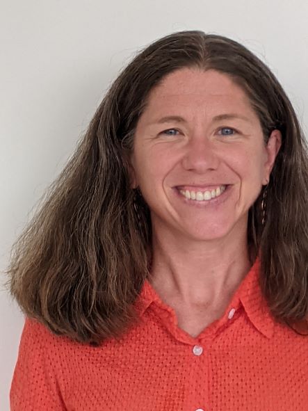
Felice Kelly: Core Scientist

Felice uses microscopy to explore the beauty of the cellular world and better understand spatially organized cellular functions. She has over a decade of experience imaging single-celled organisms and sub-cellular structures in live and fixed cells, including as a graduate student at Rockefeller University working on fission yeast, as a post-doc at Stanford studying the parasite Toxoplasma gondii, and as a senior scientist in Scott Landfear’s lab at OHSU investigating the parasite Leishmania mexicana. She particularly enjoys assisting users with high-resolution imaging (for example, protein co-localization and sub-organellar structures) but is eager to help with all of your projects. Felice splits her time between the South Waterfront and Marquam Hill campuses. When she’s not appreciating the microscopic world she can be found running or skiing through the mountains and forests of the Pacific Northwest. Contact Felice
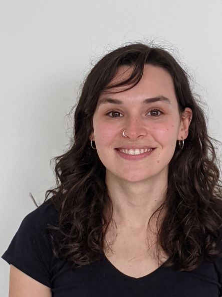
Hannah Bronstein: Associate Core Scientist

Hannah is an imaging specialist at the ALM Core. She got her first introduction to light microscopy working on her undergraduate thesis at Reed College in the Cerveny Lab using laser scanning confocal microscopy to study stem cell migration in the developing zebrafish retina. In the core, Hannah splits her time between the South Waterfront and Marquam Hill training users on a variety of light microscopes. She also offers consultation on 2D, 3D, and 4D image analysis workflows in Imaris, arivis, and ZEN. When she's not gazing through a microscope, Hannah enjoys skiing and rock climbing in the beautiful mountains of the PNW. Contact Hannah