Skull Anomaly (Craniosynostosis)
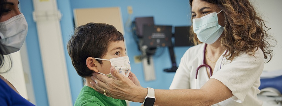
Our providers are national leaders in diagnosing and treating craniosynostosis. This condition causes an infant’s skull to grow in an unusual shape.
- We perform 60 to 80 surgeries each year, more than any other medical center in Oregon. We treat patients from throughout the west because of our expertise.
- Our team approach to care means you can see all of your child’s specialists in one day.
- Our team meets often to talk about your child’s care, making sure your child receives the best possible treatment.
- We make sure your child receives steady care over the years it takes to treat their condition. This includes regular follow-up visits for monitoring and treatment.
Understanding craniosynostosis
The word craniosynostosis comes from the Latin words for skull, together, and bones. Infants with this condition have skull bones that develop differently from other newborns. This may affect their brain development and health.
What is craniosynostosis?
When babies are born, their brains are still growing. To allow for this growth, their skulls have fontanelles (soft spots) between the skull bones. Strong, flexible tissue holds the bones of the skull together at these soft spots. These tissues are called sutures.
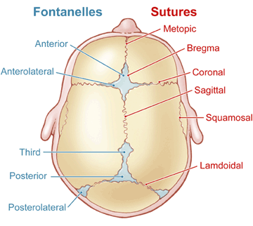
At around age 2, the skull bones join together and the sutures close. Craniosynostosis means one or more of the sutures closes early. When this happens, the brain doesn’t have room to grow. As pressure builds from the brain’s growth, the baby’s skull takes on an unusual shape.
Who gets craniosynostosis?
Craniosynostosis is rare. It affects about 4 out of 10,000 newborns in the United States, according to the Centers for Disease Control and Prevention.
What causes craniosynostosis?
We don’t know for sure what causes craniosynostosis. There are some factors that may increase your risk of having a child with craniosynostosis:
- Having thyroid disease.
- Taking certain medications, including the fertility medication clomiphene citrate, just before or early in pregnancy.
- Having a genetic condition that makes your baby more likely to have craniosynostosis involving multiple sutures.
Symptoms of craniosynostosis
Often, an unusually shaped head at birth may be the only early symptom of craniosynostosis. Other signs may appear during the first few months of your baby's life. These may include:
- Slow growth, or no growth, of your baby’s head.
- An unusual bump or shape on your baby’s skull.
- A raised, rigid line on some of your baby’s sutures.
If craniosynostosis is not treated, pressure on the brain may cause severe headaches. Over time, some children with untreated craniosynostosis may have learning disabilities or vision problems.
Screening
If your child's provider suspects craniosynostosis, they may suggest imaging tests such as X-rays or a CT scan to get a detailed look at your child’s skull. They may also refer your child to the specialists at OHSU for evaluation. Our team is skilled at identifying possible craniosynostosis based on your child’s facial features.
Types of craniosynostosis
There are different types of craniosynostosis based on its location and how many sutures are affected. There are also two categories: nonsyndromic and syndromic.
Nonsyndromic craniosynostosis
Nonsyndromic craniosynostosis is the most common type. It is not linked with any other conditions. This type typically involves the early fusion of one cranial suture. There are many subtypes:
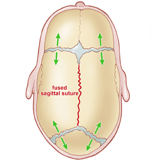
This is the most common type of craniosynostosis. It accounts for 45% of nonsyndromic cases and is usually easy to diagnose. The sagittal suture, which runs from the front to the back at the top of the skull, fuses early. The head grows long and narrow, with the back of the head becoming pointed and the forehead bulging. For most children, brain growth is normal.
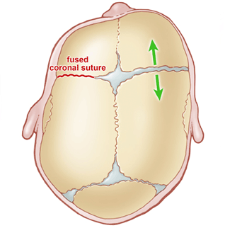
This is the second most common type of craniosynostosis. It affects the part of the skull where the forehead and the frontal lobe grow. In coronal synostosis, the forehead is flat on the affected side and bulges on the unaffected side. The nose turns toward the affected side. The eye socket is rises and tilts, giving the eye an almond shape. A child with this condition may tilt their head to avoid seeing double.
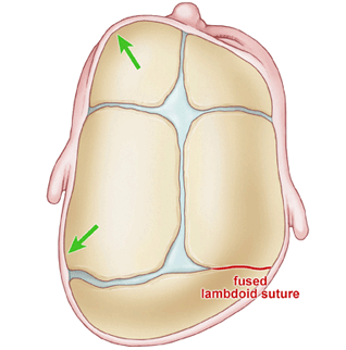
This type is rare. It happens when the lambdoid suture, which runs across the skull at the back of the head, fuses early. This may cause one side of the head to flatten, the skull to bulge behind one ear, and the affected ear to be lower. The top of the head may tilt sideways.
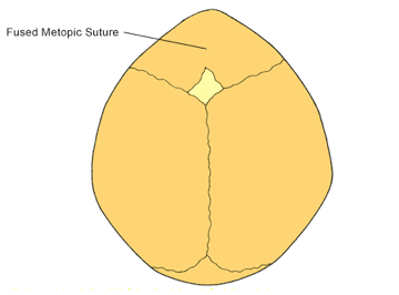
This condition is rare and it happens when a suture in the middle of the forehead fuses early. Both frontal lobes bulge forward and sideways. This gives the forehead a triangular shape and widens the back of the head. The eyes are close together and the back of the head bulges as the brain grows in that direction.
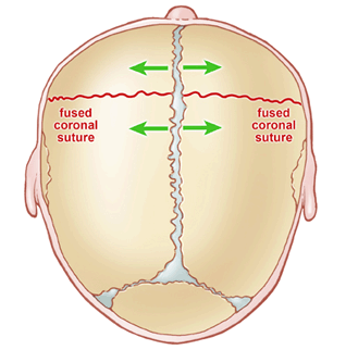
In rare cases, multiple sutures fuse early:
- Bicoronal (anterior brachycephaly): The coronal sutures, which run along the hairline on the forehead and above the ears, close early on both sides of the skull. This causes the head to grow broad and short.
- Bilambdoid (posterior brachycephaly): Both lambdoidal sutures at the back of the head close early, causing a short, wide skull.
- Sagittal plus metopic (scaphocephaly): Both the sagittal sutures at the top of the head and the metopic sutures in the middle of the forehead close early. This causes the ears to be long and narrow.
- Bicoronal, sagittal, metopic: When this group of sutures all close early, it causes the head to be short and wide or pointed at the top.
- Pancraniosynostois: The large sutures in the head fuse early. This may lead to brain pressure that can cause headaches, vision loss, seizures and developmental delays.
Syndromic craniosynostosis
In most cases, syndromic craniosynostosis is linked to a genetic condition. It can cause mild or severe developmental problems.
More than 150 syndromes are linked to craniosynostosis. The most common are Apert, Carpenter, Crouzon, Muenke, Pfeiffer and Saethre-Chotzen syndromes.
This condition leads to early closure of sutures at the top and front of the skull. It causes an unusual head shape that limits room for brain growth and the middle of the face may not develop fully. The jaws may not align, making it hard to chew or breathe during sleep. Fingers and toes may be joined or webbed. Apert syndrome may lead to developmental delays.
This condition is linked with lambdoid and sagittal synostosis. It may cause limbs to develop differently, including extra fingers or toes, or webbing on the hands or feet. Male children with Carpenter syndrome often have testicles that do not drop. The condition may lead to hearing and vision issues, heart defects and obesity.
The middle of the face and the eye sockets form differently in children with Crouzon syndrome. This affects the shape of the head, facial features and alignment of the upper and lower jaws. It can lead to difficulty breathing as the child grows. It does not usually affect intellectual development.
This genetic mutation affects bone growth. It often causes an unusual head shape, wide-set eyes and flat cheekbones. Some children with Muenke syndrome have small differences in their hands and feet, and issues with hearing or vision. About 30% have some delays in intellectual development.
This condition affects the face, hands and feet. Along with an unusual head shape, children with Pfeiffer syndrome may have shallow eye sockets, bulging eyes, short thumbs and big toes, and webbed hands or feet.
This condition affects skull growth and causes an unusual head and face shape. Children with Saethre-Chotzen syndrome may have difficulty breathing and eating. Some have learning delays, but most have normal intellectual development.
Learn more
- Craniosynostosis, Genetic and Rare Diseases Information Center
- Craniosynostosis, Centers for Disease Control and Prevention
- Support for craniofacial patients and families, Children’s Craniofacial Association
- Information and support, Faces: The National Craniofacial Association
For families
Call 503-346-0640 to:
- Request an appointment
- Seek a second opinion
- Ask a question
Locations
Parking is free for patients and their visitors.
Doernbecher Children’s Hospital
700 S.W. Campus Drive
Portland, OR 97239
Map and directions
Find other locations across Oregon and in southwest Washington.
For referring providers
Refer your patient to OHSU Doernbecher.
Call 503-346-0644 to:
- Seek provider-to-provider advice.
- Request education about plagiocephaly or other conditions.