Pediatric Kidney Disorders
Our team at OHSU Doernbecher Children’s Hospital offers advanced expertise in treating children and teens with kidney disorders.
You’ll find:
- The most complete pediatric kidney services in Oregon.
- Providers who specialize in treating children’s kidney conditions.
- The latest minimally invasive treatment options, including robot-assisted surgery.
Understanding kidney disorders
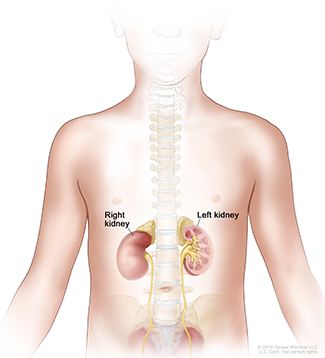
What do the kidneys do?
The kidneys are two fist-size organs just below the rib cage that:
- Filter waste and pass it as urine.
- Regulate minerals in the blood.
- Keep fluids in the body balanced.
- Produce hormones important to red blood cells, blood pressure and bone health.
Kidney anatomy
Types of kidney disorders
Most children with kidney disorders are born with them. Often, the kidneys function normally. Other times, a disorder can lead to serious health problems. With treatment, most children with kidney disorders can live full lives.
Types include:
- Congenital kidney disorders are those a child is born with.
- Acute kidney disorders, such as kidney stones, strike suddenly and are usually successfully treated.
- Chronic kidney disorders are ongoing conditions that can worsen over time.
Horseshoe kidney and pelvic kidney
Horseshoe kidney
Horseshoe kidney, also called renal fusion, is a birth defect that occurs in about 1 in 500 babies. The kidneys fuse at the lower end during fetal development, forming a U. They also may not move as high as normal kidneys.
About one-third of children with horseshoe kidney have one or more other conditions, such as fluid on the brain (hydrocephalus) or spina bifida.
Horseshoe kidneys may work normally. However, some children have complications, increasing the risk of kidney damage, such as:
- Kidney stones
- Urinary tract infections
- Swollen kidneys
- Urine backing up into the kidneys
Pelvic kidney
Pelvic kidney, a birth defect also known as ectopic kidney, happens when the kidney doesn’t move up during fetal development. A kidney can also move to the other side of the body. It affects about 1 in 1,000 children.
Many children with this condition have kidneys that work normally. Others have problems with urine flow, which can cause infections, a swollen kidney, and kidney damage. If the kidney sits especially low in the body, your child may need to wear protective gear to prevent injury during sports or other activities.
Signs of horseshoe and pelvic kidney
Horseshoe and pelvic kidney can cause complications such as urinary tract or kidney infections, kidney stones or backed-up urine. These may result in:
- Fever, chills or nausea
- Sharp pain in the back, side or lower abdomen
- Pain while urinating
- Cloudy or bloody urine
- Changes in urination frequency
Diagnosing horseshoe and pelvic kidney
Horseshoe kidney may show up on an ultrasound before your baby is born. But most of the time, these conditions aren’t found until your child has tests for something else. Tests include:
- Blood tests
- Urine tests
- Ultrasound
- X-ray with contrast dye
Learn more about our urology tests.
Treating horseshoe and pelvic kidney
Your child probably doesn’t need treatment if the kidneys work fine. In unusual cases, with kidney damage or persistent complications, we may recommend surgery.
Our pediatric urologists offer expertise in minimally invasive methods. Options include moving a pelvic kidney or moving the ureter (the tube that connects the kidney to the bladder).
Duplex kidneys and ureters
Duplex kidneys and ureters are also called a duplex collection system. One or both kidneys have two tubes (ureters), not one, draining to the bladder.
This is a birth defect and is usually found in childhood. It affects about 1% of the U.S. population, and it’s more common in girls. Causes are unclear but may be genetic.
Types of duplex kidneys and ureters
- Incomplete: The two ureters are separate leaving the kidney but join at some point and enter the bladder as one.
- Complete: The two ureters remain separate between the kidney and bladder.
Related conditions
Duplex ureters may be small or malformed, leading to complications. These can also happen in children without duplex kidneys and ureters:
- Ureterocele: The ureter’s opening into the bladder is so small that urine collects above it. The end of the ureter balloons, usually inside the bladder.
- Vesicoureteral reflux: Urine flows back into the kidney, sometimes because of a ureterocele. This may result in a kidney infection or kidney damage.
- Ectopic ureter: One of the ureters isn’t connected to the bladder properly. This can cause urine to drain outside the bladder. It usually goes into the urethra, which carries urine outside the body. In girls, it can leak into the vagina.
Signs and symptoms
If both ureters are connected to the bladder, children usually don’t show symptoms. When they do, symptoms can include:
- Back or side pain
- Dribbling or difficulty urinating
- Urinary tract infections
- Kidney infections
- High blood pressure
Diagnosing duplex kidneys and ureters
Duplex kidneys and ureters are usually found during tests for other conditions, such as an ultrasound. Your child’s doctor may recommend further tests, such as blood tests or an X-ray that uses dye put into the bladder.
Treating duplex kidneys and ureters
The kidneys and ureters themselves typically need no treatment. But complications may need treatment, such as antibiotics for infections or surgery for more serious conditions. Our pediatric urologists are experts in minimally invasive techniques such as laparoscopy, with only two small incisions.
Surgical options include:
- Endoscopy: This may be used to treat a ureterocele. A thin tube with a camera is guided through the urethra into the bladder. The balloon of urine is drained with a small incision.
- Ureteral reimplantation: The surgeon attaches an ectopic ureter to the bladder.
- Partial nephrectomy: Part of the kidney connected to an ectopic ureter is removed.
- Ureteropyelostomy: An ectopic ureter in the upper kidney is connected to the normally functioning lower kidney. Urine then flows normally through the remaining ureter.
- Flap valve: The surgeon lengthens the ureter where it connects to the bladder. This lets urine flow from the kidney but not back up into the bladder (vesicoureteral reflux).
Learn more
- Children With Chronic Kidney Disease: Tips for Parents, National Kidney Foundation
- Caring for a Child with Kidney Disease, National Institute of Diabetes and Digestive and Kidney Diseases
- Vesicoureteral Reflux (VUR) in Infants and Young Children, American Academy of Pediatrics
For families
Call 503-346-0640 to:
- Request an appointment.
- Seek a second opinion.
- Ask questions.
Locations
Parking is free for patients and their visitors.
Doernbecher Children’s Hospital
700 S.W. Campus Drive
Portland, OR 97239
Map and directions
We also offer locations in Eugene and Salem.
Refer a patient
- Refer your patient to Doernbecher.
- Call 503-346-0644 to seek provider-to-provider advice.