Body Imaging
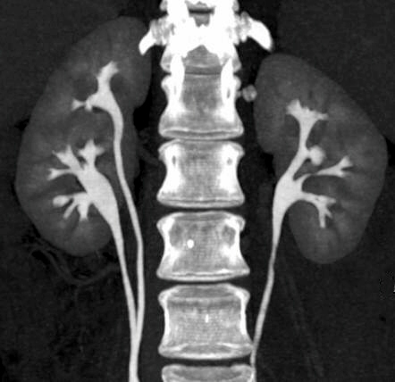
The Body Imaging section of Diagnostic Radiology provides a comprehensive range of diagnostic services at OHSU. The state of the art computed tomography (CT), magnetic resonance imaging (MRI), ultrasound, xray, and fluoroscopy services using the latest imaging techniques offer the most advanced evaluation for abdominal and pelvic pathology. The body section also performs a broad range of interventional procedures under CT or ultrasound-guidance, including solid organ biopsies and aspirations.
Each body imaging radiologist is certified by the American Board of Radiology and fellowship-trained in body imaging. The staff works closely with internal medicine, multiple surgical specialties, gynecology, and other clinical services. Weekly interdisciplinary conferences focusing on in-depth patient care are held with multiple surgical and oncologic services. Meet the Body Radiologist Team. We're expanding!
Routine and advanced imaging services provided
Dual Energy CT | CT Perfusion | CT Colonography | US Elastography | Contrast enhanced US | MRI Spectroscopy | DCE-MRI | Diffusion Weighted Imaging | Advanced Prostate MRI | Advanced Rectal MRI | MR Elastography
Our Body Radiologists can also perform US and CT guided Targeted Biopsy for Oncologic Molecular Profiling in conjunction with Knight Cancer Institute.
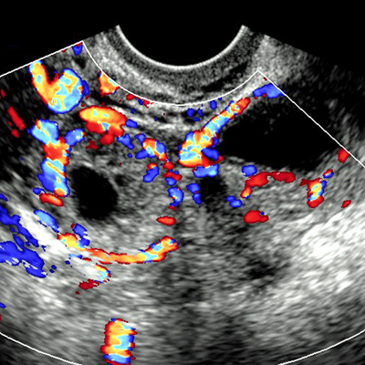
Contrast Enhanced Ultrasound (CEUS) is now offered at OHSU
CEUS is a newly offered imaging technique which makes use of tiny inert gas bubbles injected through a peripheral IV to improve US images and diagnosis. Dr. Bryan Foster, Associate Professor of Radiology and Director of Ultrasound, has been offering this new service in Diagnostic Radiology since 2017 with great success. We are currently the only institution in Oregon performing this technique and perhaps the only in the PNW.
CEUS is typically used in the characterization of liver and renal lesions and is also used in certain vascular conditions. CEUS is especially beneficial for patients with certain contrast allergies, chronic kidney disease or acute kidney injury who ordinarily cannot undergo standard contrast-enhanced CT and MRI imaging. It is also useful as alternative imaging technique to CT to minimize radiation exposure."
CEUS movie of the kidney. Split screen image with contrast image on the left and standard ultrasound on the right. Longitudinal view of the right kidney shows a small, solid, slowly-enhancing upper pole renal mass which was diagnosed as papillary renal cell carcinoma. The patient went on to partial nephrectomy and the diagnosis was confirmed on final pathology.
CEUS movie of the liver. Split screen image with contrast image on the right and standard ultrasound on the left. Transverse view of the right lobe of the liver shows a large mass with peripheral nodular enhancement and progressive central fill-in consistent with a benign liver hemangioma.
Fellowship
The Body Imaging Fellowship is a year-long commitment with concentrations in CT, MR, and ultrasound. The primary emphasis is clinical and designed to be a well-balanced training program encompassing all of the basic and advanced clinical areas of adult abdominal and pelvic imaging. The fellow receives much "hands-on" experience at the workstation as well as through active procedure services with image-guided aspirations and biopsies. Graduated autonomy and responsibility granted during the course of the year and active involvement with the residents and with interdepartmental conferences all aid in building confidence and refinement of one's skills.
The fellow will become experienced in a broad range of adult pathology with OHSU as a level 1 trauma center, an active oncology center with the Knight Cancer Institute, and a transplant center specializing in renal, pancreas, and liver. The fellow will be actively involved in protocol refinement, especially in CT and MRI. The busy ultrasound laboratory offers proficiency in obstetrics, gynecology, small parts, abdominal, and vascular exams. OHSU has state-of-the-art equipment with 10 CT scanners, one of which is a 128-slice scanner and three of which have iDose (dose reduction) capability. There are six MRI scanners, two of which are 3 tesla and 1 of which is a 1.5 tesla large bore scanner capable of scanning patients up to 500 lbs. Five ultrasounds with state of the art grayscale and Doppler capabilities are also used.
The Body Imaging Fellowship at OHSU is a non-ACGME fellowship program. Candidates must be Board Certified or Board Eligible radiologists. Candidates must also be eligible for an unrestricted Oregon state medical license prior to beginning the fellowship. Candidates for Fellowship are selected through individual applications, not through the National Residency Matching Program (NRMP).”
OHSU is located in the prestigious west hills of Portland, Oregon. Endless outdoor recreational activities are easily accessible from the city. The ocean and mountains are less than 1-1/2 hours away. Metropolitan Portland has won many awards for city planning and offers the best of urban living without sacrificing the landscape.
Application information
Our program is currently filled through June 2026. We will be recruiting for 2 positions for the 2026/27 Fellowship Year and will begin accepting applications November 1, 2024.
To apply please email:
- One completed and signed application form
- curriculum vitae
- personal statement
- three letters of recommendation
- photocopy of the Dean's letter from your medical school or ECFMG certificate
- USMLE Step 1,2,3 scores
For additional information, questions or sending completed application packets, contact Julia Starkey.
Foreign applicants must be legally able to work in the U.S., or eligible to obtain work authorization. Initial and extension J-1 Visas are considered in very rare circumstances. All non-ACGME fellowship candidates must be eligible for H-1B visas, which includes successful completion of all USMLEs.
We are unable to support J-1 applicants that require a Conrad J-1 waiver in order to be eligible for an H-1B visa. All visa applicants must have successfully passed all USMLE steps at least 6 months prior to fellowship start as USMLEs are required for visa applications. LMCC exams are not accepted for H-1B applications.