OHSU Casey Eye Institute scientists are harnessing the power of advanced imaging technologies to learn what changes may occur in the back of the eye in patients with glaucoma.
At the heart of this work is the development of Optical Coherence Tomography (OCT), a commonly used, noninvasive technique that uses light waves to capture highly detailed cross-sectional images of the inner eye. Led by David Huang, M.D., Ph.D., professor of ophthalmology and biomedical engineering, and co-inventor of OCT technology, a team of scientists at the Casey Center for Ophthalmic Optics and Lasers (COOL) lab has been using this technology to devise new ways of monitoring tissue loss in glaucoma. This includes the addition of software that allows them to map out the eye’s smallest capillaries and measure blood flow in seconds.
In collaboration with Casey Eye Institute’s Glaucoma Service, COOL lab researchers are now determining how retinal blood flow is altered in glaucoma, and how this might be improved by conventional pressure-lowering glaucoma treatments.
Recently, COOL lab ophthalmic imaging expert Yali Jia, Ph.D., and glaucoma physician-scientist John Morrison, M.D., have begun collaborating on research using an enhanced type of optical coherence tomography called visible light OCT.
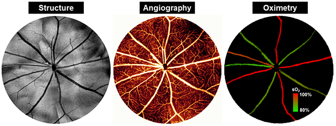
This newer OCT system, developed by Jia and her team at Casey, builds on these current imaging technologies by employing a laser with visible light to determine oxygen content within retinal blood vessels.
Currently, the researchers are refining visible light OCT technology so it is capable of measuring oxygen levels in the capillaries most likely affected by glaucoma, said Jia, associate professor of ophthalmology and biomedical engineering. Altered oxygen levels may be early signs of glaucoma’s progression and affect a patients’ susceptibility to elevated eye pressure.
“Visible light OCT provides a meaningful interpretation of the effects of glaucoma in ‘real time,’ said Morrison, professor of ophthalmology. “We hope these investigations will provide new insights into the disease and improve our ability to detect and manage glaucoma in its earliest stages, before vision is affected.”