Fine-tuning corneal procedures
OHSU Casey Eye Institute is setting a new standard in Level I evidence in corneal procedures and reducing subjectivity from treatment decisions. By embedding clinical research in patient care, OHSU Casey Eye Institute works to improve quality of life for patients.
“We know we will obtain better visual outcomes by pushing the bar with scientific investigation and utilizing new technologies,” said OHSU Casey Eye Institute ophthalmologist Winston Chamberlain, M.D., Ph.D.
Comparing corneal transplant methods
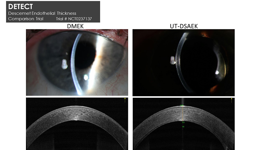
Posterior layer replacements of the cornea now account for more than 50% of corneal transplants in the U.S. In 2019, OHSU Casey Eye Institute (in collaboration the University of California, San Francisco and Stanford University) published the results of a comparison of visual outcomes and multiple other optical and surgical variables after Ultrathin Descemet stripping automated endothelial keratoplasty (UT-DSAEK) and Descemet membrane endothelial keratoplasty (DMEK). Patients included in the study had Fuchs’ endothelial dystrophy or pseudophakic bullous keratopathy. The study set a new standard for randomized, controlled trials for this corneal surgery.
“These surgeries have been challenging for corneal surgeons to learn and have rapidly evolved over the last 10 years,” said Chamberlain, who was the co-author and co-designer of the study. “It is critical to have Level 1 evidence to show we are doing
the right thing for our patients.”
The study assembled an incredibly well-matched set of patients. With randomization of treatment, Chamberlain noted there was an excellent degree of sensitivity in detecting differences.
“DMEK resulted in better vision than UT-DSAEK, with no difference in complications,” he said. “Patients’ visual acuity improved, and they recovered faster with single cell layer treatment.”
Study participants continued to have better acuity and fewer higher-order aberrations (microscopic optical distortions) in their corneas up to two years later. Data are still coming out of this trial, and the investigation has generated new questions about graft survival and the role of novel pharmacotherapeutics. OHSU Casey Eye Institute plans to pursue these questions in a larger-scale, multicenter clinical trial currently in the planning stage.
Reducing subjectivity in corneal transplant decision-making
Imaging cornea disease states is essential to staging disease and surgical intervention. Yan Li, Ph.D., of the COOL lab at OHSU Casey Eye Institute is collaborating with Chamberlain to develop a protocol to image and quantify the subtle changes in every layer of the cornea very early in corneal endothelial disease. The goal is to find thresholds of disease to guide surgical decisions for posterior layer corneal transplantation. The methods will utilize optical coherence tomography (OCT), pioneered by OHSU Casey Eye Institute’s David Huang, M.D., Ph.D.
“This has the potential to provide more objective guidelines for committing patients to surgery and also to predict optical outcomes after the surgery.” Winston Chamberlain, M.D., Ph.D.
Modifying crosslinking for better acuity
OHSU Casey Eye Institute is seeking to improve the corneal crosslinking (CXL) modality and taking the next step toward visual rehabilitation in patients with keratoconus. CXL strengthens corneal tissue by using riboflavin eye drops as a photosensitizer and ultraviolet-A. Chamberlain, Huang, Afshan Nanji, M.D., M.P.H., and Richard Stutzman, M.D., use OCT to image subtle changes in the layers of corneas affected by keratoconus. This imaging then guides the sculpting and shaping of the cornea using an excimer laser before locking in the shape with crosslinking for improved visual acuity and long-term stability.