Core Description
What is a core facility?
"Core facilities are centralized shared resources which provide access to instruments, technologies, services, as well as expert consultation and other services to scientific and clinical investigators. The typical core facility is a discrete unit within an institution and may have dedicated personnel, equipment, and space for operations. Given the rapid growth of life science research, core facilities are becoming more of a integral means to access cutting edge instrumentation and expertise. Shared resources also help foster a collaborative research environment and are an integral component of the scientific community." -NIH
What is flow cytometry?
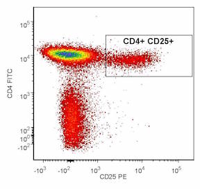
"Flow cytometry is a technology that simultaneously measures and then analyzes multiple physical characteristics of single particles, usually cells, as they flow in a fluid stream through a beam of light. The properties measured include a particle's relative size, relative granularity or internal complexity, and relative fluorescence intensity. These characteristics are determined using an optical-to-electronic coupling system that records how the cell or particle scatters incident laser light and emits fluorescence. A flow cytometer is made up of three main systems: fluidics, optics, and electronics."-BD
The OHSU Flow Cytometry Shared Resource has served OHSU researchers and the Knight Cancer Institute members since 1996 and the Oregon Stem Cell Center since 2005. Through our shared resource, researchers have access to advanced flow cytometry instrumentation, technical expertise and technical services. FCSR staff performs daily regiment of QC protocols and cleaning procedures to ensure our instruments are functioning within manufacturer recommended set of criteria.
The FCSR provides:
- Quantitative measurement of fluorescent reporters to assess the distribution of specific molecules within cell populations
- Sorting to isolate purified cell populations based on detection of specific probes such as antibodies and fluorescent proteins
- Analysis of multiple characteristics such as relative cell size, antibody binding to cell surface or intracellular biomarkers, DNA and RNA content, and fluorescent protein expression.
- Functional assays to measure apoptosis, enzyme activity or calcium flux
- Cells can be sorted into 5-15 ml conical tubes, eppendorf tubes or cloned into multi-well plates.
- After acquisition of data, investigators will receive data files and/or data plots. In addition, for sorting experiments, investigators will receive sorted cells and an analysis of post-sort purity.