AIRC 11.75T Instrument
Description
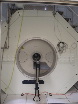
The Bruker 11.75T instrument is located in the Lamfrom Biomedical Research Building. The 11.75 Tesla (T) horizontal magnet has a clear bore diameter of 31 cm and the ID of the large gradient set is 20 cm. Higher gradient strengths and faster slew rates are available from a gradient insert that has an 9 cm ID. The RF transmitter unit contains two broadband frequency synthesizers and two digital RF transmitters capable of producing 180-600 MHz and 6‑365 MHz pulses, respectively, with each amplifier capable of ≥1 kW peak power. This instrument is equipped with a Resonance Research, Inc high-bandwidth shim power supply for demanding quantitative imaging and spectroscopy studies.
Technical Information
- Field Strength/1H Frequency: 11.75 Tesla (T)/500 MHz
- Clear bore/Working bore [insert]: 31 cm/20 cm [9 cm]
- Gradient Strength/Slew Rate [20 cm insert]: ≥200 mT/m/ ≥1000 T/(m*s)
- Gradient Strength/Slew Rate [9 cm insert]: ≥750 mT/m/≥7500 T/(m*s)
- Transmit channels: 2
- Receiver channels: 2
- Length/Mass: 2.5m/12 tons (60 tons with iron shield)
Radio Frequency Coils
The system is equipped with two actively RF-decoupled 1H volume resonators with ID/OD of 7.2/8.9 cm and 15.4/19.7 cm, respectively, a 1H 2 cm diameter surface receiver coil, and a 1H/31P double-tuned mini surface coil.
Accessories
- SA Instruments Inc animal monitoring and gating system (model 1025)
- Ventilator (model CWE)
- Gas anesthesia
MR Instruments
- Siemens 3T Prisma
- Siemens 3T Trio (ONPRC)
- Siemens MAGNETOM 7T
- Bruker 11.75T
Sample Images
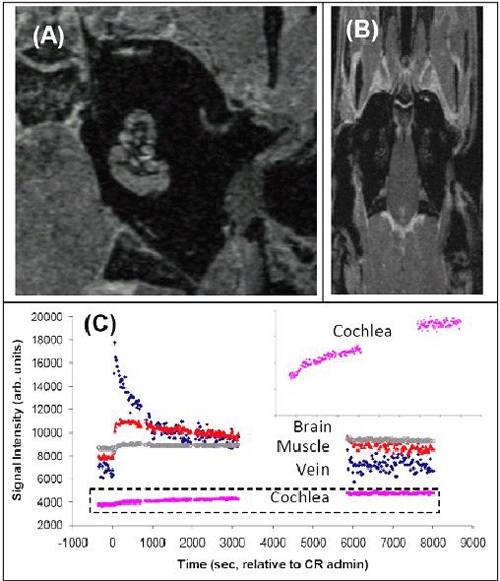
In vivo guinea pig MRI acquired at 12 T. In panel (A) is a 98 μm isotropic anatomical image showing the left cochlear of the live guinea pig. In panel (B) is a two-dimension T1-weighted gradient recalled echo image (100 μm in-plane 500 μm through-plane) collected as part of a dynamic contrast enhancement study to investigate cochlea perfusion. In this case a low-molecular weight gadolinium based contrast reagent (CR) was used. In panel (C) is shown signal intensity time course for different tissue regions of the DCE study. Significant CR uptake is observed for the four tissues investigated. (Selzer, Omelchenko, Nuttall, Rooney).
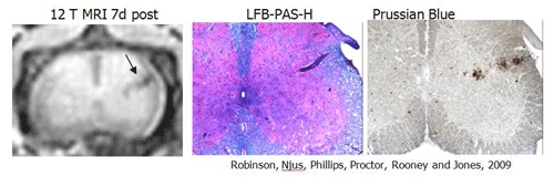
Iron labeled T-cell tracking at 12T. Feridex labelled T-cells were used to induce experiement allergic encephalomyelits in the mouse via adoptive transfer. The panel at the left shows a 12T T2-weighted MRI of the mouse spinal cord acquired 7 days post adoptive transfer. The large hypointense T2-weighted MRI areas indicated by the arrow corresponds to demyelination and iron accumulation as indicated by luxol-fast blue and Prussian blue coregistered histological sections.