Brain Tumor Symptoms and Diagnosis
At the OHSU Brain Institute, our doctors can analyze the genetic features of tumors. This helps us choose the right treatment for you. We offer you:
- Expert pathologists who analyze tissue samples. Our neuropathologists specialize in brain tumor diagnosis.
- An in-house lab where we identify tumor traits for precise, personal treatment.
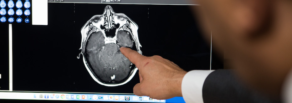
- High-resolution MRIs to learn the tumor’s size, location and more so we can plan the best treatment.
- Fast appointments, usually within two weeks of your referral.
- A weekly tumor board meeting where experts from many specialties work together on the next steps in your care.
Brain tumor symptoms
Brain tumor symptoms are similar to those of many other conditions. Our first step is to find if your symptoms have a different cause.
Symptoms include:
- Headaches
- Seizures
- Hearing or vision loss
- Double vision
- Fatigue
- Depression or behavioral changes
- Cognitive changes, including trouble thinking or speaking
- Weakness or paralysis on one side of the body
- Loss of balance or dizziness
- Nausea or vomiting
- Facial numbness
Diagnosing brain tumors
We sometimes find brain tumors when otherwise healthy people suddenly have a seizure, unusual weakness or speech problems. If you have these symptoms, go to an emergency room. If ER doctors suspect a brain tumor, they may send you to us for a full evaluation.
Other times, tumors are found on a brain scan done for another reason, such as to check for a concussion. Or you might go to your regular care provider with more subtle symptoms. Your provider may send you to us for more testing if they have concerns.
We use advanced technology for a precise diagnosis. This gives your care team the information they need to create the right treatment plan for you. Learn more about our brain tumor treatment options.
Exam
We’ll start with a physical exam and ask about your medical history. We’ll review previous scans, tests and treatments.
Imaging
Imaging scans give us detailed pictures of a tumor’s size and location. The radiologists who read and analyze your images are brain tumor specialists.
MRI: Magnetic resonance imaging uses radio waves and powerful magnets to create detailed images of the tumor and surrounding brain areas.
- Mapping: We often use MRI to map your brain before surgery.
- Contrast agents: Advanced software and new types of contrast agents (special dyes) highlight problem areas.
Our MRI tools include:
iMRI: We may use MRI during surgery (intraoperative) to protect healthy tissue and make sure we’ve removed as much of the tumor as possible. We have the only iMRI scanner in Oregon. Our scanner, in OHSU Doernbecher Children’s Hospital, is available for adults too.
PET-MRI: This combines positron emission tomography and MRI for detailed images. Few centers have this technology. We use a safe amount of a radioactive substance to track the tumor’s development or to see if medications are reaching the tumor. This helps us see which medications work best.
MRA and MRV: Magnetic resonance angiography and venography show the brain’s blood vessels. Neurosurgeons may use these to map your brain before surgery.
MRS: Magnetic resonance spectroscopy measures biochemical changes in the brain. It helps us learn the tumor type.
Perfusion MRI: A contrast dye helps us see the amount of blood going to different parts of the brain. This helps doctors decide where to do a biopsy (removing a piece of tumor for analysis).
fMRI: A functional MRI helps doctors see which parts of the brain handle functions such as speech, sensation and movement. Doctors use fMRI so they know which areas to avoid during surgery or radiation therapy.
Diffusion tensor imaging: By mapping how water travels through the brain, doctors can protect blood vessels during surgery.
Other imaging tools:
CT scan: Computed tomography is a powerful X-ray that can provide detailed images of the tumor. Doctors use CT scans for brain tumors if MRI isn’t an option.
PET scan: We inject a small and safe amount of a radioactive substance to highlight where a tumor is growing.
Biopsy
In a biopsy, we remove a piece of tumor for analysis. In some cases, a sample is taken at the same time as surgery to remove the tumor.
- You receive anesthesia so you don’t feel pain.
- We remove a piece or all of the tumor.
- Expert pathologists analyze the tumor tissue under a microscope to determine the type of tumor and how aggressive it is.
- Pathologists may do other tests, such as looking for any genetic mutations that match with treatments.
- Full results are available within a few days. After we confirm your diagnosis, we can plan the next steps in your care.
We remove tissue using these methods:
Craniotomy (open biopsy): The neurosurgeon temporarily removes a piece of skull to reach all or most of the tumor. At OHSU, we are experts in awake craniotomies. This method protects brain function. During surgery, our team can test your functions by asking you to speak or move. Our pathologists may analyze tissue during surgery to guide the procedure.
Stereotactic needle biopsy: We may use this method if a craniotomy is too risky. We use computer-assisted imaging to map the brain and locate the best place for the incision. We make a small hole in the skull and use imaging to guide a needle to the tumor so we can extract a sample.
Lumbar puncture
This procedure is also called a spinal tap. We withdraw cerebrospinal fluid (CSF) to look for cancer cells. CSF, a clear liquid, bathes the brain and spinal cord to cushion them, bring nutrients and remove waste. During a lumbar puncture:
- You lie on your side.
- We use a needle to withdraw fluid from between the bones in your spine.
- We send the fluid to our on-site lab for fast, precise analysis.
Blood and urine tests
We do not use blood or urine tests to diagnose brain tumors. However, we may use them to check how well your organs are working, especially if you will have surgery.
Learn more
- Signs and Symptoms of Adult Brain and Spinal Cord Tumors, American Cancer Society
- Brain Tumor: Symptoms and Signs, Cancer.net
For patients
- Referral: To become a patient, please ask your doctor for a referral.
- Questions: For questions or follow-up appointments, call 503-494-5626.
Location
Parking is free for patients and their visitors.
OHSU Neuro-Oncology Clinic, Marquam Hill
3270 SW Pavilion Loop, 2nd floor
Portland, OR 97239
Map and directions
Refer a patient
- Refer your patient to OHSU.
- Call 503-494-4567 to seek provider-to-provider advice.
Patient resources
Imaging tumors, improving outcomes
Neuroradiologist Ramon Barajas focuses on improving techniques to measure brain tumors.