MR Temporal Arteritis - Vasculitis - Brain WWO
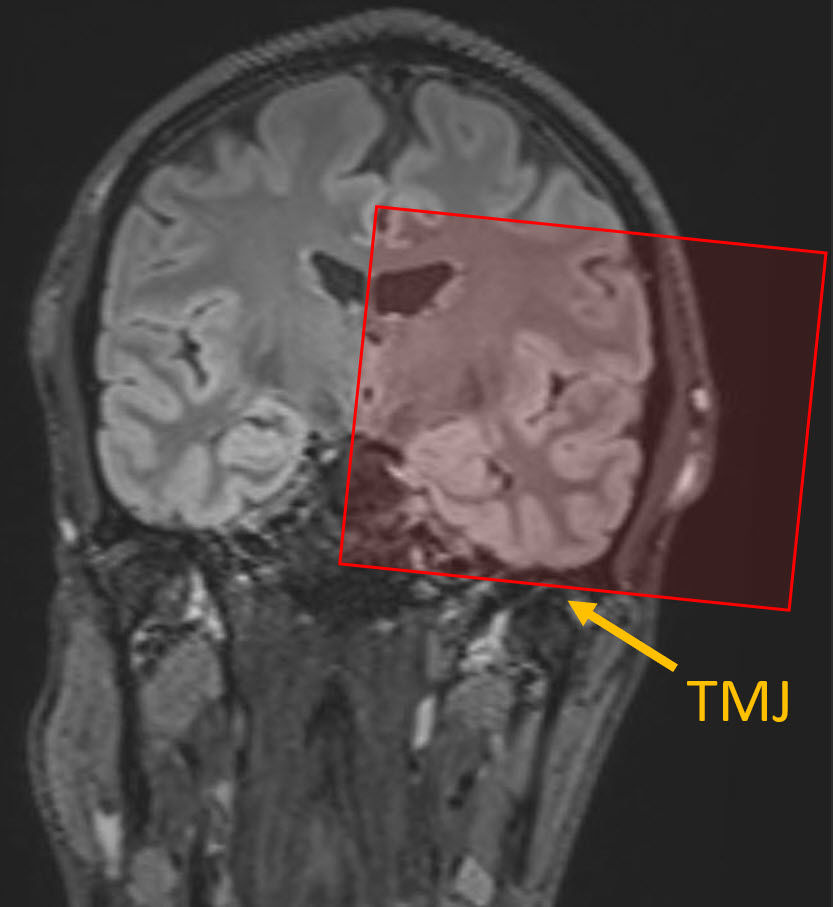
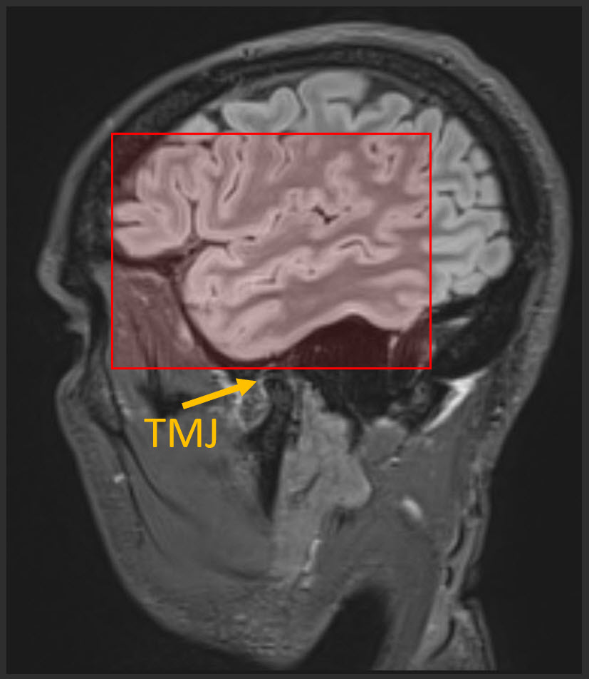
Coverage on VWI sequences:
- Run separate sequences for right and left scalp.
- Keep FOV 10 x 10 cm
- Inferior-most slice in stack should be centered on the TMJ (see pic below).
- 22-26 slices
Other notes:
- Scanner preference: MR3 only.
- Coil: 64-channel if patient can tolerate.
- Last updated: 11/11/2023
- Charge as: Brain WWO & MRA Brain WO
| Plane | Weighting | Mode | Slice (mm) | Gap (mm) | FAT SAT | FOV (cm) | MPR | Notes |
|---|---|---|---|---|---|---|---|---|
| AXIAL | DWI | EPI | 3 | 0.3 | YES | 23 | no | Angle to Corpus. Cover Skull Base to Vertex. Send only B1000 & ADC. |
| AXIAL | TOF | 3D FFE | 1 | 0 | no | 20 | MIP COW, Right, Left and Posterior | |
| SAG | FLAIR | 3D IR-TSE | 1 | 0 | YES | 23 | no | NO ANGLE. Cover ears and nose. Spacing and gap are variable. |
| SAG | T1 | 3D TSE | 1 | 0 | NO | 23 | no | NO ANGLE. Cover ears and nose. Spacing and gap are variable. |
| INJECT CONTRAST | ||||||||
| SAG | T1 | 3D TSE | 1 | 0 | YES | 23 | AXIAL, COR | NO ANGLE. Cover ears and nose. Spacing and gap are variable. |
| AXIAL-Right | T1-VWI | 2D TSE | 3 | 1 | YES | 10 | no | Right scalp - see coverage note above. |
| AXIAL-Left | T1-VWI | 2D TSE | 3 | 1 | YES | 10 | no | Left scalp - see coverage note above. |