MR CONGENITAL FEMALE PELVIS WO BODY Protocol
Scan Notes
Last updated: 10/30/23
Charge as: Pelvis WO
- Send ADC maps.
- Void before exam.
Breath Holds
- Scan on expiration.
- Monitor that patient is breath-holding. Breathe the patient slowly so they have time to follow instructions. Do not start scan until the patient has stopped breathing.
- Give 2L O2 if it will help with breath-holds UNLESS PATIENT HAS COPD OR ANOTHER REASON NOT TO GIVE O2.
| Plane | Weighting | Mode | SLICE | GAP | FAT SAT | FOV | SCAN RANGE | Notes |
|---|---|---|---|---|---|---|---|---|
| COR | T2 | SSTSE BH | 4 mm | 0 mm | N | Top of kidneys ? pelvis | Sacrum ? anterior abdominal wall | Scan sacrum to anterior abdominal wall. CONFIRM GOOD COIL PLACEMENT. Large FOV to include kidneys. Pelvic pathology is often related to renal pathology. |
| SAG | T2 | TSE | 4 mm | 0 mm | N | 24 mm/ Fit to Patient | Mid-femoral head ? mid-femoral head | Scan mid-femoral head to mid-femoral head. Freq A-P. Consider using an anterior Sat band if lots of abdominal wall motion. |
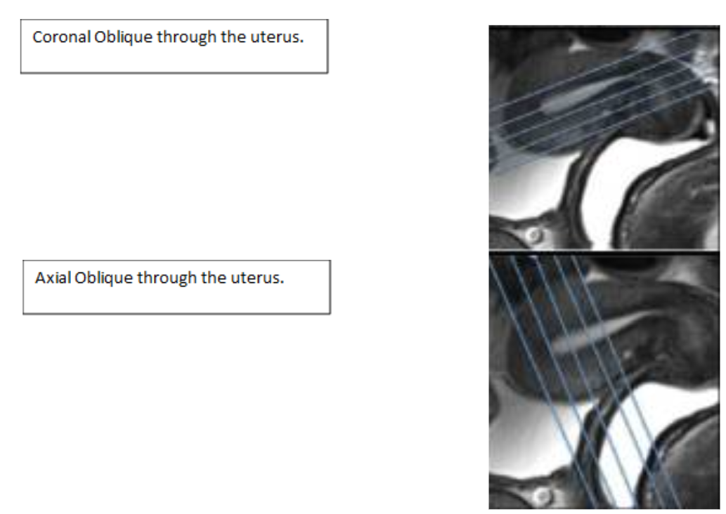
| AX OBLIQUE | T2 | TSE | 4 mm | 0 mm | N | 20-24 mm/ Fit to Patient | Uterus ? perineum | Scan uterus to perineum.?Slices should be along the length of the uterus. CALL Rad FOR PLANNING! |
| COR OBLIQUE | T2 | TSE | 4 mm | 0 mm | N | 20-24 mm/Fit to Patient | Include Uterus, cervix, and vagina | Include Uterus, Cervix and vagina.?Slices should be along the short axis of the uterus. CALL Rad FOR PLANNING! |
| AX | T1 | 3D THRIVE HIGH RESOLUTION pre | -- | -- | Y | Match AX TSE T2 Pelvis | Match AX TSE T2 Pelvis | HIGH RESOLUTION THRIVEs. |
| AX | T2 | DWI | 5 mm | 1 mm | SPIR | Match AX TSE T2 Pelvis | Match AX TSE T2 Pelvis | Trigger & track. Free-breathing sequence, so please position slices accordingly. B=0, 500, 1000. |